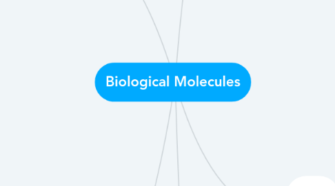
1. Carbohydrates
1.1. Monosaccharides
1.1.1. Single sugars, monomers, make carbs
1.1.2. Trioses, pentoses, hexoses, isomers
1.1.3. Benedicts test for reducing sugars
1.1.3.1. Benedicts Reagent turns from blue to red when heated with a reducing sugar
1.1.3.2. Colorimetry- the greater the absorbance the less glucose in solution
1.2. Disaccharides + polysaccharides
1.2.1. Di - when 2 monosaccharide molecules bond - condensation reaction, forms water - glycosidic bond
1.2.1.1. Sucrose = alpha glucose + beta fructose
1.2.1.2. Testing for non-reducing sugars - if doesn't test positive with benedicts reagent. HEAT solution with dilute hydrochloric acid, then test with benedicts test if positive then was a non-reducing sugar.
1.2.2. Poly - many monosaccharides joined together with condensation reactions e.g. starch
1.2.2.1. Testing for starch - add water, add iodine - solution will turn blue/black
1.2.3. Disaccharide examples
1.2.3.1. MALTOSE = glucose+glucose
1.2.3.2. SUCROSE = glucose+fructose
1.2.3.3. LACTOSE = glucose+galactose
2. Lipids
2.1. Triglycerides
2.1.1. Ester of fatty acids and glycerol
2.1.2. Condensation reaction
2.1.3. 3 molecules of fatty acid + 1 molecule of glycerol -> triglyceride + 3 molecules of h2o
2.2. Structure + function
2.2.1. Insoluble - good energy storage molecules
2.2.1.1. Readily hydrolysed to fatty acids and glycerol
2.2.2. Readily hydrolysed to fatty acids and glycerol
2.2.3. Can pass through phospholipid bilayer
2.3. Roles
2.3.1. Energy source - insoluble in water
2.3.2. Insulation - fat=poor conductor of heat, fatty myelin sheath surrounding neurons prevents voltage loss from axons
2.3.3. Protection - protects delicate organs (kidneys)
2.3.4. Structural - phospholipid bilayer important
2.4. Phospholipids
2.4.1. 2 fatty acid molecules, 1 phosphate group, 1 glycerol
2.4.2. Phosphate group = hydrophilic. Fatty acids = hydrophobic
2.5. Emulsion test
2.5.1. Add ethanol, shake, add equal volume of water, shake, milky white EMULSION if positive
3. Intro
3.1. Molarity
3.1.1. Mass of substance/ molar mass of substance
3.2. Chemical Bonds
3.2.1. Ionic
3.2.1.1. The electrostatic force between ions holds them together
3.2.2. Covalent
3.2.2.1. Held together by sharing a pair of electrons in their outer shells
3.2.3. Hydrogen
3.2.3.1. Forms when electrons are shared unequally between atoms - polar molecule (hydrophilic)
3.3. Condensation Reactions
3.3.1. Join together monomers to form macromolecules - this produces a molecule of water. Reverse is hydrolysis.
4. Starch, Glycogen, Cellulose
4.1. Starch
4.1.1. Polysaccharide formed from condensation reactions between molecules of alpha glucose
4.1.1.1. AMYLOSE
4.1.1.1.1. Unbranched chains - coil into a helical structure - glycosidic bonds
4.1.1.1.2. Dense can be packed into a small space - not soluble good for storage
4.1.1.2. AMYLOPECTIN
4.1.1.2.1. Branched chains with glycosidic bonds
4.2. Glycogen
4.2.1. Like AMYLOPECTIN
4.2.1.1. Highly branched - less dense and more soluble than starch
4.3. Cellulose
4.3.1. Polysaccharide formed from condensation reactions between beta glucose.
4.3.2. Hydrogen bonds cross link chains into bundles called microfibrils
4.3.3. Further hydrogen bonding binds microfibrils to fibres
4.4. Food Stores
4.4.1. Enzyme-catalysed hydrolyses break down starch and glycogen into glucose
4.4.2. When glucose is oxidised during respiration energy is RELEASED so cells can synthesise ATP. Condensation reactions polymerise glucose forming starch + glycogen
4.4.3. Bc Starch + glycogen both insoluble don't affect osmotic properties so can be stored in cells. Starch = plant, Glycogen = animal
5. Proteins
5.1. Amino acids
5.1.1. Each have a diff R group - there are 20 diff R groups
5.2. Peptide bonds
5.2.1. Carboxyl group of 1 amino acid joins to the amino group of another, molecule of water formed (condensation reaction). Peptide bond forms
5.3. Protein structure
5.3.1. Primary structure
5.3.1.1. the sequence of amino acids, hydrogen bonds form
5.3.2. Secondary structure
5.3.2.1. hydrogen bonding causes the chain to coil into an alpha helix or beta pleated sheet (depends where bonds are)
5.3.3. Tertiary structre
5.3.3.1. hydrogen, ionic and disulphide bonds form - weak bonds so pH and temp affect structure
5.3.4. Quaternary structure
5.3.4.1. when a protein consists of 2 or more polypeptide chains e.g. haemoglobin
5.4. Biuret Test
5.4.1. Add alkaline solution of copper sulphate - turn pink/purple
