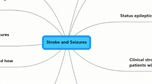
Stroke and Seizures
Door Paul de Roos


1. 1. Structure of talk
2. Thrombolysis and seizures
2.1. picture
2.2. picture
2.2.1. recurrence rate is lower than in other treatment modalities
2.3. picture
2.3.1. Seizure incidence and haematoma location
2.3.1.1. Frontal region highest prevalence
2.3.2. Status epilepticus is more frequent
2.3.2.1. picture missed
3. Non-convulsive inhibitory seizures
3.1. picture
3.2. picture
3.3. picture
4. Are stroke-related seizures harmful?
4.1. YES
4.2. picture
4.3. picture
4.4. picture
5. To treat? and how
5.1. picture
5.2. picture
6. Clinical stroke types in patients with seizures
6.1. Ischaemic seizures
6.1.1. picture
6.2. Clinical stroke types
6.2.1. pictures
6.3. Locations of stroke
6.3.1. pictures
6.4. Vascular risk factors
6.4.1. picture
6.5. Post stroke EEG patterns
6.5.1. picture
6.6. Recurrence according to time of onset
6.6.1. picture
6.7. Median NIHS Scores in the three seizure groups
6.7.1. picture
6.8. Median mRankin Scores in the Three Seizure groups
6.8.1. picture
7. Author
7.1. J. De Reuck
8. 2. Review of literature
8.1. Reference
8.1.1. Bladin et al, Arch Neurol, 2000
8.2. picture
8.2.1. picture
8.2.1.1. picture
9. 2. Classification
9.1. Old classification
9.1.1. early onset
9.1.1.1. 95%
9.1.1.1.1. within 48%
9.1.2. Late onset
9.1.2.1. months after stroke
9.1.2.2. Stroke types in very late onset
9.1.2.2.1. picture
9.1.2.2.2. Lacunar stroke
9.2. New proposed classification
9.2.1. picture
10. Status epilepticus
10.1. picture
10.2. picture
10.3. picture
10.4. status epilepticus and infarct
10.4.1. temporal lobe neocortex
10.4.1.1. picture
