

1. Signs and Symptoms
1.1. Numbness/weakness in face, arm or leg
1.2. Facial drooping on one side when smiling
1.3. Slurred speech (aphasia)
1.4. Sudden vision problems
1.5. Dizziness, loss of balance
1.6. Difficulty swallowing (dysphagia)
1.7. Sudden severe headache, especially with hemorrhagic stroke
1.8. Confusion
2. Body System Impacts
2.1. Respiratory System
2.1.1. Aspiration Pneumonia from impaired swallowing
2.2. Heart/ blood vessels
2.2.1. DVT, arrhythmias
2.3. Neurological System
2.3.1. Paralysis, sensory loss, memory issues, seizures, cognitive deficits
2.4. Muscular system
2.4.1. Weakness, spasticity, fall risk, muscle atrophy
2.5. Gastrointestinal and urinary systems
2.5.1. Bowel/Bladder incontinence, UTIs from catheter use
2.6. Endocrine & Metabolic
2.6.1. poor temperature and glucose regulation
2.7. Psychological
2.7.1. Depression, anxiety, emotional instability.
3. Complications
3.1. Brain swelling (Cerebral edema) leading to Increased Intracranial pressure
3.2. Seizures
3.3. Recurrent strokes or heart attacks
3.4. Recurrent strokes or heart attacks
3.5. PE or DVT
4. Diagnostic test
4.1. Imaging Studies
4.1.1. Non-contrast CT scan of the head
4.1.1.1. First line test to rule out hemorrhagic stroke
4.1.1.1.1. May show ischemic changes after a few hours
4.1.2. MRI of the brain
4.1.2.1. More sensitive than CT for early ischemic stroke
4.1.2.1.1. Identifies location and extent of brain injury
4.1.3. CT Angiography CTA/MR Angiography (MRA)
4.1.3.1. Detects blood vessel occlusions, aneurysms, or vascular malformations
4.1.4. Carotid Doppler Ultrasound
4.1.4.1. Evaluates carotid artery stenosis (common source of emboli)
4.1.5. Echocardiogram (especially transesophageal)
4.1.5.1. Identifies cardiac sources of emboli (e.g., atrial thrombus, valve disease)
4.2. Laboratory Tests
4.2.1. Complete Blood Count, PT, APTT, INR, Blood glucose, Serum electrolyte, BUN and Creatine, Lipid profile, Troponin
4.2.2. Toxicology screen
4.2.3. Cardiac & Rhythm Tests: ECG ( to detect AF and other arrhythmias)
4.2.4. Holter monitor or Telemetry for continuous monitoring for intermittent arrhythmias
5. Treatment Options
5.1. Acute phase: Ischemic Stroke
5.1.1. IV Thrombolysis (tPA)
5.1.1.1. Alteplase: 0.9mg/kg (max 90mg) within 3-4.5 hours of symptom onset
5.1.1.2. Tenecteplase: Bolus alternative
5.1.2. Endovascular Mechanical Thrombectomy
5.1.2.1. For large vessel occlusions within 6 hours (possibly up to 24hrs with penumbra imaging.
5.1.3. Early Antiplatelet Therapy (if not receiving tPA)
5.1.3.1. Aspirin 150-300 mg within 24-48hrs
5.2. Acute Phase: Hemorrhagic Stroke
5.2.1. Blood Pressure Management
5.2.1.1. IV agents (e.g Labetalol, nicardipine) to keep SBP<140-160mmhg
5.2.2. Neurosurgical referral
5.2.2.1. Neurosurgery/neurovascular consult for clot evacuation or aneurysm intervention
5.3. Acute Support & Secondary Prevention
5.3.1. Cardiac Workup & Monitoring
5.3.1.1. ECG, telemetry to detect AF (initiate OACs like warfarin or DOACs)
5.3.2. Vascular Interventions
5.3.2.1. Carotid endarterectomy or stenting if significant carotid stenosis identified
5.3.2.2. Carotid endarterectomy or stenting if significant carotid stenosis identified
5.3.3. Lipid Lowering Agents (Statins) and blood pressure control agents
5.4. . Rehabilitation & Referrals
5.4.1. Physical therapy
5.4.1.1. Early mobilization to improve strength, balance, prevent complications
5.4.2. Occupational Therapy
5.4.2.1. Assist in regaining ADL skills, promote independence
5.4.3. Speech and Language Therapy
5.4.3.1. For aphasia, dysarthria, swallowing issues
5.4.4. Neuropsychological support
5.4.4.1. Address cognitive deficits, emotional health, mood disorders
5.4.5. Specialized Interventions
5.4.5.1. Neurostimulation (tDCS) and Botulinum toxin injections for spasticity management
5.4.6. Referral to neurology/Stroke Clinic
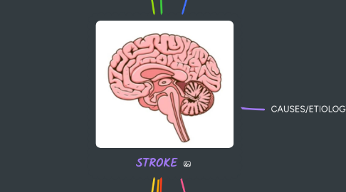
6. DEFINITION AND PATHOPHYSIOLOGY
6.1. sudden interruption of blood supply to the brain, leading to brain cell damage due to lack of oxygen and nutrients.
6.2. Ischemic stroke (most common, 87%)
6.2.1. (a) Caused by interruption of cerebral blood flow due to
6.2.1.1. Thrombosis (blood clot forms in the brain)
6.2.1.2. Embolism (clot travels from another part of the body, often the heart) lead to
6.2.2. (b) Decreased oxygen/glucose delivery
6.2.3. (C) Cellular hypoxia and death - Infarction
6.2.4. (d) Penumbra zone: area of potentially salvageable brain tissue
6.2.5. (d) Inflammatory cascade- further damage
6.3. Hemorrhagic stroke
6.3.1. (a) Caused by rupture of blood vessel- bleeding into brain tissue
6.3.2. (b) caused by Hypertension, Aneurysm rupture, Arteriovenous malformation (AVM)
6.3.3. (c) Results in Increased Intracranial pressure ICP
6.3.4. (d) Compression of brain tissue
6.3.5. (e) Rapid neurological deterioration
6.4. (4) Transient Ischemic Attack (TIA) Mini stroke
6.4.1. Temporary blockage <24 hours
6.4.1.1. Warning sign of full stroke
7. CAUSES/ETIOLOGY
7.1. Ischemic Stroke
7.1.1. Atherosclerosis, AF, Carotid artery stenosis, Hypercoagulable states e.g cancer
7.2. Hemorrhagic stroke
7.2.1. Chronic hypertension, head trauma, Blood vessel malformations, Anticoagulant therapy (increased bleeding risk)
8. RISK FACTOR
8.1. 1. Non -Modifiable
8.1.1. Age (increased risk>55 years)
8.1.2. Male gender
8.1.3. Family history of stroke or cardiovascular disease
8.1.4. Ethnicity (e.g, African American higher risk)
8.2. 2. Modifiable
8.2.1. Hypertension (leading cause)
8.2.2. Smoking (vasoconstriction and atherosclerosis)
8.2.3. Diabetes mellitus (Vascular damage)
8.2.4. High cholesterol (atherosclerosis)
8.2.5. AF and other heart diseases
8.2.6. Obesity and Physical inactivity
8.2.7. Excessive alcohol use
8.2.8. Drug use (e.g. cocaine, amphetamines)