
1. Differential
1.1. Functionality
1.1.1. Functional (20%)
1.1.1.1. Hypercortisolism (Cushing syndrome) 7%
1.1.1.1.1. Presentation: Weight gain, moon facies, buffalo hump, central obesity, proximal muscle weakness (difficulty standing from seated position or climbing stairs), poor wound healing → striae + easy bruising + plethora, hirsutism, amenorrhea, depression, HTN, DM, polyuria (from hypokalemia), osteoporosis, psychiatric sx
1.1.1.2. Hyperaldosteronism (Conn syndrome) 1%
1.1.1.2.1. Presentation: Muscle cramps, weakness, HTN
1.1.1.3. Catecholamine Hypersecretion (NE & E) = pheochromocytoma 5%
1.1.1.3.1. Presentation: Episodic tachycardia, sustained or episodic HTN, HA, flushing, palpitations [classic triad = HA, flushing, palpitations]
1.1.1.4. Androgen hypersecretion <1%
1.1.1.4.1. Presentation: Virilization, hirsutism, menstrual abnormalities
1.1.2. Non-functional
1.1.2.1. Benign (majority; 80%)
1.1.2.1.1. Nonfunctional adenoma
1.1.2.1.2. Myelolipoma
1.1.2.1.3. Cyst
1.1.2.1.4. Hematoma
1.1.2.1.5. Granuloma/infection
1.2. Malignant potential
1.2.1. Benign
1.2.2. Malignant
1.2.2.1. Adrenocortical carcinoma (May be a functional -oma!) 5%
1.2.2.1.1. Notes: • Lethal; 5-yr survival < 25% • 60% functional; 75% > 6 cm at presentation • Most common hypersecretion: Cushing syndrome > Minors [Androgens > Feminization] • Presentation: tumor-specific hypersecretion; abdominal mass, pain, nausea, early satiety, anorexia, wt loss.
1.2.2.2. Metaastasis of other primary tumors (most common: lung & breast) 2%
1.2.2.2.1. Note: If pt has hx of extra-adrenal tumor, prevalence of metastasis is up to 70%
2. Work-up
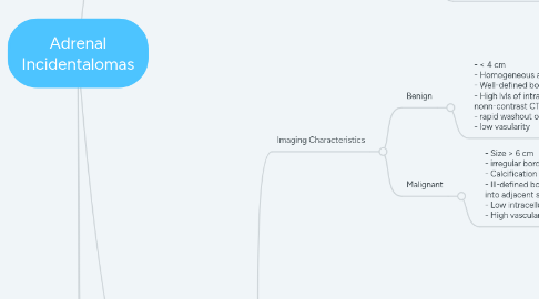
2.1. Imaging Characteristics
2.1.1. Benign
2.1.1.1. - < 4 cm - Homogeneous appearance - Well-defined borders - High lvls of intracellular lipid (<10 HU on nonn-contrast CT) - rapid washout of contrast - low vasularity
2.1.2. Malignant
2.1.2.1. - Size > 6 cm - irregular borders w/ necrosis - Calcification +/- hemorrhage w/i the mass - Ill-defined borderes w/ possible invasion into adjacent structures - Low intracellular lipid - High vascularity
2.2. Adrenal mass discovered on imaging
2.2.1. H&P (include medication hx; seek for endogenous source of glucocorticoids, including those contained in herbal/alternative supplements
2.2.1.1. Lab testing for hormone hypersecretion
2.2.1.1.1. Suspected Hypercortisolism
2.2.1.1.2. Suspected Pheochromocytoma
2.2.1.1.3. HTN
2.2.1.1.4. If no clinical sx are present, send all the above biochemical studies
3. Treatment
3.1. Functional adrenal mass
3.1.1. Adrenalectomy regardless of size (as long as pt is medically fit to undergo operation)
3.2. Non-functional mass
3.2.1. Size impacts management:
3.2.1.1. < 4 cm w/ benign features (< 5% malignancy
3.2.1.1.1. Observe w/ interval CT scanning
3.2.1.2. 4-6 cm
3.2.1.2.1. Observation or resection is acceptable - decide based on patient-specific factors s.a. lvl of perioperative risk
3.2.1.3. > 6 cm w/ concerning features
3.2.1.3.1. Laparoscopic adrenalectomy