

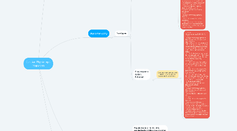
1. Autorhmycity
1.1. Two types
1.1.1. Fast response Action Potential
1.1.1.1. Generated automatic cells Ex: SA node and AV node
1.1.1.1.1. PHASE 4 - Unstable membrane RMP due to the decreased permeability of the membrane to K+. This would cause the K+ to decreased influx of K+ making the membrane negative (-60mV). PHASE 0 - Involved: Na+ leaks and slow voltage-gated Ca2+ channels - The cell becomes more permeable to Na+, thus the leaky Na+ channels will allow Na+ to go inside the cell, making the cell more positive, until it reached the critical firing level or -40mV. - At -40mV: slow voltage-gated Ca2+ channels will open -> Na+ and Ca2+ going inside the cell -> more positive until it reaches the peak to become Phase 1 or 2 PHASE 1or 2 - Involved: slow voltage-gated K+ channels and slow voltage-gated Ca2+ channels - At the peak: slow voltage-gated K+ channels will open -> K+ efflux-> cell becomes more negative because of the efflux of the positive ions -> slow because of the long-lasting Ca2+ channels - At the end of phase 2: The Ca2+ channels will close, so the K+channels are the ones that are left opened. PHASE 3 - Involved: slow voltage-gated K+ channels - Efflux of K+ causes the cell to depolarize. - Hyperpolarization: K+ channels are opened at an extended amount of time ->more positive ions go out -> more negative
1.1.2. Slow response Action Potential
1.1.2.1. Generated non-automatic cells Ex: Atrial muscle, Ventricular muscles and Purkinje Fibers
1.1.2.1.1. PHASE 4 - Stable membrane RMP: -90mV PHASE 0 - Involved: fast voltage-gated Na+ channels and slow voltage-gated Ca2+ channels - The cell becomes more permeable to K+, and slightly to Na+ -> the cell becomes more positive. Some slow voltage-gated Na+ channels open ->Na+ influx -> until it reaches -65mV - At -65mV: The fast voltage-gated Na+ channels will open -> Na+ influx -> rapid depolarization, but before the membrane completely depolarizes. - At -20mV: The slow long-lasting voltage-gated Ca2+ channels will open -> until it reaches the spike PHASE1 - Involved: slow voltage-gated K+ channels and slow voltage-gated Ca2+ channels - At the spike: fast voltage-gated Na+ channels will close, and the slow voltage-gated K+ channels will open -> K+ efflux ---> making the membrane more positive - The slow voltage-gated Ca2+ channels are still open PHASE 2 - Involved: slow voltage-gated K+ channels and slow voltage-gated Ca2+ channels - Plateau: Equal K+ efflux and Ca2+ influx PHASE 3 - Involved: slow voltage-gated K+ channels - Ca2+ channels will close -> allowing K+ efflux to dominate -> making the cell more negative due to the efflux of positive ions - Final repolarization and no hyperpolarization because the K+ channels are opened long enough so when it reaches the bottom they are already closed.
2. Contractility
2.1. Positive intropic effect or increase contractility
2.1.1. o Increased Heart Rate o Sympathetic via beta 1 Receptors catecholamines o Cardiac Glycoside o High Extracellular Ca o Low Extracellular Na o Left Ventricular size
2.2. Negative intropic effect or decrease contractility
2.2.1. o Parasympathetic stimulation o Heart Failure o Myocardial infarction o Pharmacologic depressants o Hypoxemia, Hypercarbia acidosis o Ca channel blockers o High extracellular Na
3. Conductivity
3.1. Repolarization - In the atria, repolarization follow the direction of depolarization: o SA node -> AV node o The first part to depolarize is the first to repolarize. o The last part to depolarize is the last to repolarize. - In the ventricles, the repolarization occurs in the opposite direction of the depolarization: o Postero-basal -> apex -> antero-basal o The first part to depolarize is the last part to repolarize o The last part to depolarize is the first part to repolarize.
3.2. Depolarization: Antero-basal -> SA node -> 3 internodal tracts -> AV node -> bundle of His -> left to right bundle branches -> Purkinje fiber @ the cardiac apex -> postero-basal
4. Excitability
4.1. The cardiac muscle cells is surrounded by a membrane called sarcolemma, the invagination of the membrane will form the T tubule.
4.1.1. Structures in the sarcolemma: 1. Sodium-Potassium pump – pumps 3 Na+ out, in exchange for 2 K+ in. This is the primary active transport. 2. Sodium-Calcium exchanger – pumps 3 Na+ ions in, in exchange for 1 Ca2+ out. This is secondary active transport. 3. Calcium pump – that will actively transport out. This is primary active transport
4.1.1.1. Structures in the T tubules: 1. Sodium-Calcium exchanger 2. Voltage-gated Calcium channels – the receptors are dihydropyridine receptors (DHP).
4.1.1.1.1. Structures inside the cell 1. Myofilaments – contains the contractile proteins 2. Sarcoplasmic reticulum a. Ryanodine receptors (RYR) –found on the membrane of SR; these are Calcium-gated Ca2+ channels, these will open when Ca2+ from the ECF binds to it. b. Calcium pump – found on the membrane; actively transport calcium back. 3. Phospholamban – inhibit the Ca2+ pump