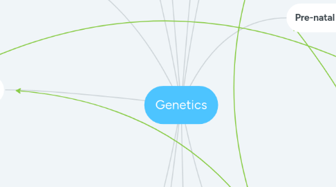
1. Chromosomes
1.1. Somatic cells (body cells): contain the normal number of chromosomes called the diploid number (ex: skin cells, brain cells)
1.2. Gametic cells (sex cells): reproductive cells that contain half the normal amount of chromosomes, called the haploid number (ex: sperm cells, eggs)
1.2.1. Male gamete: Sperm produced in testes. meiosis in males = spermatogenesis (sperm)
1.2.1.1. XY chromosome
1.2.2. Female gamete: Ovum produced in ovaries. Meiosis in females = oogenesis (ova)
1.2.2.1. XX chromosome
1.3. Structure of chromosome: centromere (holds 2 chromatids together), gene (segment of DNA that codes for a trait) and chromatids (identical copies)
1.4. Karyotype: Display of an individuals chromosomes that determines genetic disorders such as down syndrome
1.5. Homologous chromosomes: pair of chromosomes that are similar in shape and size (humans have 23 pairs. 22 autosomes and 1 pair sex, either XX or XY
2. Meiosis |
2.1. Interphase |: Chromosomes replicate, each duplicates
2.2. Meiosis |: Cell division that reduces chromosome number by half. Includes 4 phases
2.2.1. Prophase |: longest and most complex phase. 90% of the process is spent in this phase. Chromosomes condense, synapsis occurs, crossing over occurs
2.2.2. Metaphase |: shortest phase, tetrads align in the middle, independent assortment occurs, orientation of pairs to poles is random
2.2.3. Anaphase |: Sister chromatids remain attached at centromeres and homologous move towards poles (seperation)
2.2.4. Telophase |: Final phase, each pole has haploid set of chromosomes, cytokinesis occurs and 2 haploid daughter cells form
2.3. Genetic variation occurs because of crossing over and independent assortment. The seperation and assortment of the homologous chromosomes when they are divided is random, and when crossing over occurs, the swapping of genetic material between chromosomes results in new allele combinations in daughter cells
2.4. This process is used to create gametes (sex cells). It happens in two phases: Meiosis | and meiosis ||
3. Patterns of reproduction
3.1. Genotype: genetic makeup of an organism. Includes alleles that code for a trait (represented by capital and lowercase letters)
3.1.1. Alleles: variations of a physical trait
3.1.1.1. Dominant allele: described using capital letters, this trait is always expressed
3.1.1.2. Recessive allele: described using lowercase letters, this trait is only expressed when not paired with a dominant allele
3.1.1.2.1. Principal of dominance: Developed by Mendel, this theory states in crossbreeding organisms, the dominant trait always is expressed over the recessive
3.1.1.3. Hybrid/heterozygous alleles: 2 different alleles for a trait
3.1.1.4. Pure/homozygous alleles: same alleles for a trait
3.2. Phenotype: appearance of a trait in an organism (physically)
3.3. Punnet squares: used to calculate the genotypes and phenotypes of the next generation
3.4. Monohybrid cross: determining the possible offspring for one trait. When both parents are heterozygous, the phenotypic ratio will always be 3:1
3.5. Dihybrid cross: determining the possible offspring for two traits. When both parents are heterozygous, according to the Mendelian ratio, it will always have a phenotypic ratio of 9:3:3:1
4. Non-disjunction
4.1. Non-disjunction is when chromosomes don't divide correctly in meiosis and results in chromosomal abnormalities that can cause disorders such as Down's Syndrome.
4.1.1. It is more common in older people
4.2. Chromosomal abnormalities usually occur in anaphase when they are not pulled to the correct side of the cell (anaphase |) or one of the chromosomes failed to separate into 2 chromatids (anaphase ||).
4.2.1. It is better to happen in meiosis || because then the child only has a 50% chance of having issues (only half have incorrect number as opposed to all daughter cells if the error occurs in meiosis |)
5. Incomplete and co-dominance
5.1. Incomplete dominance: every genotype has it's own phenotype (one allele not completely dominant over the other phenotype). Third phenotype that is blending parental traits.
5.1.1. Ex: RR = Red WW = White RW = Pink
5.2. Co-dominance: neither allele is dominant, both are expressed. (cross between both phenotypes that are both shown).
5.2.1. Ex: Black chicken x white chicken -> speckled chicken with black and white feathers
6. Meiosis ||
6.1. Meiosis ||: No interphase, no more DNA replication and all phases are the same as mitosis
6.1.1. Prophase ||: chromatin coils to chromosomes, centrioles repel and form spindle fibres, nuclear membrane and nucleus break down
6.1.2. Metaphase ||: chromosomes line up along centre line
6.1.3. Anaphase ||: sister chromatids separate and go to opposite poles
6.1.4. Telophase ||: daughter chromosomes reattach, cytokinesis occurs (final division), 4 haploid daughter cells produced (gametes)
7. DNA
7.1. Contains all of our genetic information in a ladder-like structure called the double helix
7.2. DNA molecules made up of nucleotides: Phosphate group, pentose sugar, nitrogenous bases
7.2.1. Phosphate group and pentose sugar form backbone of DNA molecule and the bases form the "rungs" of the ladder
7.2.2. 4 types of nitrogenous bases. Adenine, guanine, thymine, cytosine (AT form base pair and GC)
7.2.2.1. These bases are arranged in triplets called codons
7.2.2.1.1. Stop codons: sequence transition ends
7.2.2.1.2. Start codons: Protein sequence transition begins
7.3. Gene: Section of DNA that codes for protein
7.3.1. DNA -> Gene -> protein -> physical trait
8. RNA
8.1. DNA never leaves the nucleus, so to send information to the rest of the body, it uses a messenger - mRNA
8.2. Differs from DNA in 3 ways: single stranded vs double stranded, ribose sugar vs deoxyribose, contains uracil (U) vs thymine (T)
9. Pre-natal genetic testing
9.1. Amniocentesis: procedure that detects/rules out fetal health issues. Doctor takes amniotic fluid sample that has cells (genetic information). Usually done between 15-18 weeks in the pregnancy
9.2. Chorionic villus sampling (CVS): Done within weeks 10-14, this takes. sample of placenta with fetal cells inside. It can be done via needle through abdomen or through vagina. Risks include cramping, infection, pre-term labour or miscarriage
9.3. Nuchal translucency scan: 11-14 weeks, done transabdonminally, scans for genetic abnormality and structural problems
9.4. Genetic counselling: Helps you understand the genetic risks for your child, tests and what your results mean
9.5. Down's syndrome: extra copy of chromosome 21. Kleinfelters syndrome: extra sex chromosome. Turner's syndrome: missing sex chromosome
10. Reproductive strategies & technologies
10.1. Artificial insemination: Sperm is collected and inserted in a woman's vagina
10.2. In Vitro fertilization (IVF): eggs and sperm are collected and made in a lab and then inserted in a uterus
10.2.1. Preimplantation genetic diagnoses: Genetic testing on an embryo in the lab before implantation
10.3. STEM cells: undifferentiated cell that still has potential to specialize in the body
10.3.1. Embryonic: obtained from embryos
10.3.2. Adult: somatic cells that have retained the ability to specialize
10.3.3. Induced puriponent: specialized adult cells that have been induced to return to a STEM-like-state
10.3.4. Can help treat diseases like cancer and diabetes and solves the problem of tissue rejection because it is a genetic match to the patient
10.4. Transgenic organisms: organisms whose genetic material includes DNA from different species
10.4.1. Can increase nutrient contents in foods, produces insulin after being modified and transgenic animals can serve as organ donors for humans or produce milk with human proteins
10.4.2. Risks/issues: 1. Environmental threats - genes may create superweeds/bugs by crossing to other species 2. Health - long-term effects unknown 3. Social/economic - research costs a lot and there are ethical debates of modifying and using these species
