

1. Parts of the Neuron
1.1. Dendrites
1.1.1. Dendrite: A short branched extension of a nerve cell
1.2. Soma/Cell bodies
1.2.1. Cell body: Core region contains the nucleus, integrates the signals coming into the dendrites and has dendritic spines
1.3. Axon
1.3.1. Axon: Carries information to be passed onto other cells, may branch to reach out and contact many other neurons
1.4. Presynaptic terminals
1.4.1. Presynaptic terminals: Where neurotransmitters are packaged into vesicles
1.4.1.1. Synaptic vesicles: “Sacks” of neurotransmitter contained in the axon terminals of the pre-synaptic cell
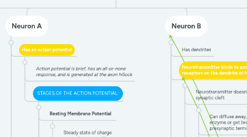
2. Neuron A
2.1. Has an action potential
2.1.1. Action potential is brief, has an all-or-none response, and is generated at the axon hillock
2.1.2. STAGES OF THE ACTION POTENTIAL:
2.1.2.1. Resting Membrane Potential
2.1.2.1.1. Steady state of charge separation across the membrane, approximately -70 mV
2.1.2.2. Threshold of Activation
2.1.2.2.1. Neuron reaches action potential
2.1.2.3. Depolarization
2.1.2.3.1. The cell becomes more POSITIVE
2.1.2.3.2. The resting membrane potential is becoming more positive and the action potential is traveling down the axon
2.1.2.3.3. INFLUX of cations OR EFFLUX of anions
2.1.2.4. Overshoot
2.1.2.4.1. Peak
2.1.2.4.2. Membrane potential is positive
2.1.2.5. Repolarization
2.1.2.5.1. The cell becomes more NEGATIVE
2.1.2.5.2. Voltage-gated channels open, falling phase
2.1.2.5.3. INFLUX of anions OR EFFLUX of cations
2.1.2.6. Hyper polarization
2.1.2.6.1. The cell becomes more negative, it is further from threshold potential
2.2. The action potential travels down the axon
2.3. Voltage-gated Calcium (Ca2+) channels open
2.4. Creates influx of Ca2+
2.4.1. Ca2+ enters the presynaptic terminal
2.5. Ca2+ causes vesicular fusion
2.5.1. Vesicles release the neurotransmitter
2.6. Neurotransmitter is released into the synaptic cleft
2.6.1. Neurotransmitter (NT): A chemical substance released by a pre-synaptic neuron to communicate with a post-synaptic neuron
2.6.2. Can be Classical or Non-Classical
2.7. Action potential reaches the axon terminal where the axon from Neuron A synapses on the dendrite from Neuron B
3. Neuron B
3.1. Has dendrites
3.2. Neurotransmitter binds to postsynaptic receptors on the dendrite of Neuron B
3.2.1. Neurotransmitter doesn't stay in the synaptic cleft
3.2.1.1. Can diffuse away, get broken down by an enzyme or get brought back to the presynaptic terminal
3.2.1.2. Neuron B has an action potential
3.2.2. CLASSES OF RECEPTORS:
3.2.2.1. Ionotropic Receptors
3.2.2.1.1. FAST-ACTING NEUROTRANSMISSION
3.2.2.1.2. The neurotransmitter binds to a receptor which is a ligand gated ion channel
3.2.2.2. Metabotropic Receptors
3.2.2.2.1. SLOW-ACTING NEUROTRANSMISSION
3.2.2.2.2. Act slowly but response lasts longer
3.2.2.2.3. Work by activating G proteins
3.2.2.3. Excitation/Inhibition of the post-synaptic neuron depends on the RECEPTOR
3.2.3. Na+/K+ Pump
3.2.3.1. Na+/K+ Pump: Actively maintains the concentration of Na+ and K+ across the cell membrane
3.2.3.2. In Neuron B, at rest, voltage-gated Na+ and K+ channels are closed
3.2.3.3. Some K+ is constantly leaking out of the membrane pores that stay open
3.2.3.4. If there are enough EPSP's in the dendrites and cell body of Neuron B, the membrane potential is raised to threshold potential
3.2.4. At threshold potential, ligand-gated Ion channels open, letting in Na+ ions
3.2.4.1. Ion channel: A protein embedded in the cell membrane that allows the passage of ion
3.2.4.2. Ligand-gated channel: Open or close only when a specific substance binds
3.2.4.3. Voltage-gated channel: Open or close only at specific membrane voltages
3.3. Influx of Na+ depolarizes the Neuron
3.3.1. Positively charged Na+ ions rush into the cell causing depolarization
3.3.2. Neuron B is closer to threshold potential
3.3.3. Receives EPSP's
3.3.3.1. EPSP: Excitatory postsynaptic potential
3.3.3.2. Summation of EPSP's raise the membrane potential to threshold potential
3.3.3.3. These bind to receptors that depolarize the neuron
3.4. Neuron B reaches overshoot
3.4.1. Voltage-gated K+ channels open and Na+ channels close
3.5. Efflux of K+ repolarizes the Neuron
3.5.1. Cell becomes more negative and moves further from threshold potential
3.5.2. K+ ions exit through voltage-gated ion channels
3.6. Because more K+ channels are open than at rest, this hyper polarizes the neuron and the membrane potential becomes more negative than at rest
3.7. When the voltage-gated K+ channels close, resting membrane potential is restored
4. Neuron C
4.1. Neurotransmitter is released at the synapse on Neuron C after Neuron B's action potential moved down the axon
4.2. Has receptors to open a Cl- channel
4.2.1. Cl- moves into the dendrite and causes an IPSP
4.3. Receives IPSP's
4.3.1. IPSP: Inhibitory postsynaptic potential