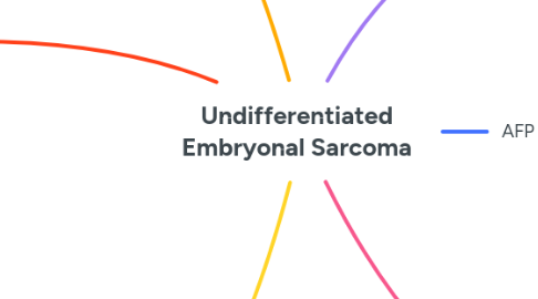
Undifferentiated Embryonal Sarcoma
da Devyani Bhatt


1. AFP Normal
2. Demographics
2.1. 6-10 years old
2.2. slight M>F
3. MR
3.1. CSF attenuation
3.2. T1
3.2.1. low
3.3. T2
3.3.1. high
3.4. useful findings
3.4.1. low intensity fibrous capsule
3.4.2. debris/fluid fluid level
3.4.3. septae
3.4.4. haemorrhage
3.5. MR used to assess resectability
3.5.1. venous structures
3.5.2. biliary tree
3.5.3. lymph nodes
4. Tx and Prognosis
4.1. resect
4.2. chemo before hand to shrink tumour
4.3. prognosis is better with multimodal tx
4.4. mets to
4.4.1. lung
4.4.2. pleura
4.4.3. peritoneum
5. USS
5.1. looks solid
5.2. iso/hyperchoic to normal liver
5.2.1. with small hypoechoic spaces
5.2.1.1. necrosis
5.2.1.2. cystic degeneration
5.2.1.3. old haemorrhage
6. CT
6.1. looks cystic
6.2. foci of soft tissue
6.2.1. peripheries
6.2.2. or forming septae
6.3. enhancement
6.3.1. peripheral on delayed imaging
6.3.2. dense, enhancing peripheral rim
6.3.2.1. pseudocapsule
