
1. Digestive System
2. Diagnosis
2.1. Constipation
2.2. Disturbed body image
2.3. Bowel incontinence
2.4. Diarrhea
2.5. Nausea
2.6. Deficit knowledge (nutrition)
3. Planning
3.1. Patient establishes a regular defecation schedule.
3.2. Patient lists proper fluid and food intake needed to soften stool and promote regular bowel elimination.
3.3. Patient implements a regular exercise program.
3.4. Patient reports daily passage of soft, formed brown stool.
3.5. Patient does not report straining or discomfort associated with defecation.
4. Implementation
4.1. Prevention: Screening for Colorectal Cancer
4.1.1. 50 years +, will need a colonoscopy every 10 years
4.2. Maintain privacy during defacation
4.3. Stool softeners to help make the stool softer for when the time comes to have a BM they won't strain
4.4. laxatives for short term use
4.5. Antidiarrheals
4.6. Enemas: promote defacation by stimulating peristalsis
4.7. Digital removal of stool: break up the hardened stool that's impacted and bring it out one section at a time
4.8. Nasogastric tube: used for decompression of abdomen Removes secretions and gaseous substances from gastrointestinal (GI) tract; prevention or relief of abdominal distention
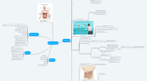
5. Assessment
5.1. Healthy Bowel Movement
5.1.1. Physical Assessment
5.1.1.1. Mouth
5.1.1.1.1. Inspect the patient’s teeth, tongue and gums.
5.1.1.2. Abdomen
5.1.1.2.1. 1. Inspect all four abdominal quadrants. 2. Auscultate the abdomen with a stethoscope to assess bowel sounds in each quadrant. 3. Percussion - identify underlying abdominal structures and detect lesions, fluid, or gas within the abdomen. 4. Palpate the abdomen for masses or areas of tenderness.
5.1.1.3. Rectum
5.1.1.3.1. Inspect the area around the anus for lesions, discoloration, inflammation, and hemorrhoids.
5.1.2. No straining
5.1.3. Don't need meds to have a bowel movement
5.1.4. No urgency
5.1.5. No blood in stool
5.1.6. Bowel Sounds Present in all 4 Quadrants
5.1.7. No Pain (no straining, no pain on palpation)
5.1.8. Frequency: Varies but 1-3 days is acceptable. Ask about their last bowel movement....
5.1.9. Color/Consistency: For infants it's soft, yellow for the older adults it's brown, formed
5.1.10. Flatulence: Accumulation of gas in the intestines causing the walls to stretch -encourage ambulation to promote the passage of flatulence
5.2. Factors Affecting Bowel Elimination
5.2.1. Age
5.2.1.1. Infants: breast milk stools, watery/yellow-brown
5.2.1.2. Toddlers: Bowel control 2-3 yrs old
5.2.2. Diet
5.2.2.1. Fiber intake
5.2.2.2. Foods that increase gas: cauliflower, apples
5.2.2.3. Foods that have a laxative effect: figs, prunes, chocolate
5.2.2.4. Foods that increase risk for constipation: pasta, cheeses
5.2.3. Fluid Intake
5.2.4. Physical Activity
5.2.4.1. stimulates intestinal activity
5.2.4.1.1. going for a walk after surgery, will help bowels start moving to get the gas moving
5.2.5. Psychosocial factors
5.2.5.1. Emotional distress increases peristalsis
5.2.5.2. Depression can decrease peristalsis, increases your patient's risk for constipation
5.2.6. Personal habits
5.2.6.1. No privacy in the hospital
5.2.7. Positioning
5.2.7.1. Normal: squatting position
5.2.7.2. Immobility: lying supine to use the bedpan
5.2.8. Pregnancy
5.2.8.1. slower peristalsis
5.2.9. Pain
5.2.9.1. Are they in too much pain to get up and used the bathroom?
5.2.9.2. Opioid use can cause constipation
5.2.10. Surgery/Anesthesia
5.2.10.1. Temporarily slows down intestinal activity
5.2.10.1.1. This is the reason why we auscultate bowel sounds before advancing their diet
5.2.11. Medications
5.2.11.1. Certain antibiotics can cause loose stools/diarrhea
5.2.11.2. Laxatives: chronic use of them can cause constipation because it weakens the bowels, can actually cause constipation
5.2.12. Hemorrhoids
5.2.12.1. Engorged, dilated blood vessels in the rectal wall
5.2.12.2. Causes: pregnancy, difficulty defacating (straining)
5.2.12.3. Symptoms: itchy, painful, bloody after defacation
5.2.12.4. Ice packs, sitz baths, ointments
5.3. Constipation
5.3.1. Impaction
5.3.1.1. Results from unrelieved constipation; a collection of hardened feces wedged in the rectum that a person cannot expel
5.3.1.2. continuous oozing of liquid stool occurs
5.4. Diarrhea
5.4.1. Incontinence
5.4.1.1. Inability to control passage of feces and gas to the anus
5.4.2. Clostridium difficile (C. difficile)
5.4.2.1. symptoms range from mild diarrhea to severe colitis
5.4.3. Inflammatory Bowel Disease (Chrohn's disease & Ulcerative Colitis)
5.5. Labs/Diagnostics
5.5.1. Monitor Hemoglobin & Hematocrit (may show anemia from GI bleed)
5.5.2. Guaiac Fecal Occult Blood Test
5.5.2.1. measures microscopic amounts of blood in the feces
5.5.2.1.1. Primary prevention tool that's used for colorectal cancer screening
5.5.3. ELISA – detects C. difficile A & B
5.5.4. Diagnostics
5.5.4.1. Radiologic
5.5.4.1.1. Direct Visualization
5.5.4.1.2. Indirect Visualization
5.6. Bowel Diversions
5.6.1. Surgically created in the abdominal wall to let fecal matter pass
5.6.1.1. temporary or permanent openings (stoma)
5.6.2. Colostomy ends in the colon (Sigmoid Colostomy)
5.6.2.1. More formed stools
5.6.3. Ileostomy ends in the ileum
5.6.3.1. Creates frequent, liquid stools (the stool leaves the body before encountering the colon, i.e. large intestine)
5.6.3.1.1. What does the large intestine absorb? What will this do to our patient?
5.6.4. Transverse colostomy
5.6.4.1. Loop colostomies are reversible stomas that may be constructed in the ileum or the colon.
5.6.4.2. thick liquid to soft consistency stools
5.6.4.3. The surgeon pulls a loop of intestine onto the abdomen and may place a plastic rod, bridge, or rubber catheter temporarily under the bowel loop to keep it from slipping back. The surgeon then opens the bowel and sutures it to the skin of the abdomen. The loop ostomy has two openings through the stoma. The proximal end drains fecal effluent, and the distal portion drains mucus.
5.6.5. End Colostomy
5.6.5.1. The end colostomy consists of a stoma formed by bringing a piece of intestine out through a surgically created opening in the abdominal wall, turning it down like a turtleneck and suturing it to the abdominal wall. The intestine distal to the stoma is either removed or sewn closed and left in the abdominal cavity.
5.6.5.2. End ostomies may be permanent or reversible. The rectum may be left intact or removed.
5.6.6. Ileoanal pouch anastomosis
5.6.6.1. 1. Colon is removed 2. pouch created from the end of the small intestine, and attaches the pouch to the patient’s anus.
5.6.6.1.1. The pouch collects fecal matter, so it stimulates the stool to come out of the anus
5.6.7. Kock Pouch
5.6.7.1. Continent Ileostomy
5.6.7.2. The pouch is created from small intestine
5.6.7.3. The pouch has a continent stoma on the abdomen created with a valve that can be drained only when the patient places a large catheter into the stoma. The patient empties the pouch several times a day.