
1. Signalment and History
1.1. cough
1.2. respiratory distress
1.3. regurgitation
1.4. trauma
1.5. Breed or species prone to thoracic disease
1.6. Metastasis screening
1.7. geriatric screening
2. Technically acceptable
2.1. Correct views
2.2. Positioned appropriately
2.3. Proper exposure
2.4. Inspiration
3. Physical exam
3.1. Signs of primary intrathoracic disease
3.2. abnormal heart or lung sounds
3.3. palpable abnormality
3.4. systemic disease
3.4.1. fever
3.4.2. sepsis
3.4.3. immune-mediated disease
3.4.4. paraneoplastic syndrome
4. Problem List
5. Evaluate body wall
5.1. skeletal structures
5.1.1. fracture
5.1.2. aggressive bone lesion(s)
5.1.3. malformation
5.1.4. degenerative change
5.2. diaphragm
5.2.1. normal appearance
5.2.1.1. diaphragmatic hernia
5.2.1.2. peritoneopericardial diaphragmatic hernia
5.3. soft tissues
5.3.1. thickening with soft tissue opacity
5.3.1.1. mass
5.3.2. thickening with gas
5.3.2.1. abscess
5.3.2.2. wound
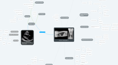
6. Thoracic point of care ultrasound
6.1. Evaluate pleural space
6.1.1. absent glide sign
6.1.1.1. pneumothorax
6.1.2. pleural effusion
6.2. Evaluate cardiac structures
6.2.1. pericardial effusion
6.3. Evaluate lungs
6.3.1. b-lines
6.3.1.1. "wet lung"
6.3.1.1.1. pulmonary edema
6.3.1.1.2. early pneumonia
6.3.1.1.3. few with atelectasis
6.3.2. consolidation
6.3.2.1. pneumonia
6.3.2.2. neoplasia
7. Other diagnostics
7.1. POC bloodwork
7.2. Referral images
7.3. Urinalysis
8. Evaluate lungs
8.1. Under inflated lungs
8.1.1. Too white
8.1.1.1. Atelectasis
8.2. Well inflated lungs
8.2.1. Too white
8.2.1.1. unstructured
8.2.1.1.1. alveolar (consolidation)
8.2.1.1.2. interstitial (ground glass)
8.2.1.2. structured
8.2.1.2.1. Nodules/masses (dots)
8.2.1.2.2. Bronchovascular (lines and rings)
8.3. Hyperinflated lungs
8.3.1. Too black
8.3.2. Too white
8.3.2.1. Bronchovascular (lines and rings)
9. Evaluate pleural space
9.1. lungs don't reach the periphery of the thoracic cavity
9.1.1. soft tissue opacity in the pleural space
9.1.1.1. pleural effusion or mass/nodules
9.1.1.2. diaphragmatic hernia (see body wall)
9.1.2. Gas opacity in the pleural space
9.1.2.1. pneumothorax
9.1.3. fat opacity in the pleural space
9.1.3.1. incomplete pulmonary expansion
10. Evaluate cardiovascular structures
10.1. Size of cardiac silhouette
10.1.1. small
10.1.1.1. hypovolemia
10.1.2. normal
10.1.3. enlarged
10.1.3.1. left-sided enlargement
10.1.3.2. generalized
10.1.3.3. globoid
10.1.3.4. right-sided enlargement
10.2. Pulmonary blood vessels
10.2.1. small
10.2.2. normal
10.2.3. enlarged veins
10.2.4. enlarged arteries
11. Evaluate mediastinum
11.1. cranial mediastinum
11.1.1. normal
11.1.2. wide
11.1.2.1. fat deposition
11.1.2.2. soft tissue opaque mass
11.1.2.3. enlarged esophagus
11.2. esophagus
11.2.1. segmental enlargement
11.2.1.1. foreign body?
11.2.2. diffuse enlargement
11.2.3. normal (not seen)
11.3. lymph nodes
11.3.1. normal (not seen)
11.3.2. enlarged, soft tissue opaque nodules/masses
11.4. trachea
11.4.1. normal diameter
11.4.2. narrow
11.5. Can you see serosal margins of mediastinal structures, including the great vessels, esophagus, and trachea?
11.5.1. Pneumomediastinum
12. Evaluate extra-thoracic anatomy
12.1. visible portion of abdomen
12.1.1. poor serosal detail
12.1.2. liver abnormalities
12.1.3. gastric abnormalities
12.2. limbs
12.2.1. aggressive bone lesion(s)
12.2.2. degenerative joint disease
12.2.3. trauma