1. Sporulation Structures
1.1. Conidia
1.1.1. Conidiophores
1.2. Sporangiospores
1.2.1. Sporangium
1.2.2. Sac-like
2. Mold
2.1. Grow @ 25C
2.2. Multicellular organism
2.3. Filamentous form
2.3.1. Fuzzy growth
2.3.2. Velvety growth
2.4. Reproduce
2.4.1. Spores
2.4.2. Conidia
2.5. Colorful cells
3. Eukaryotic organism
3.1. Membrane bound organelles
4. Transmission
4.1. Inhale spores
4.2. Environmental exposure
4.3. Tissue trauma (puncture)
4.4. Person to person
4.4.1. Extremely rare
5. Fungal Identification
5.1. Clinical information
5.1.1. Site location
5.1.2. Appearance of lesion
5.1.3. Geographic location
5.1.4. Travel history
5.2. Culture characteristics
5.2.1. Surface texture
5.2.2. Pigmentation
5.3. Microscopic evaluation
5.3.1. Elements
5.3.1.1. Conidia
5.3.1.1.1. Size
5.3.1.1.2. Free flowing spores or seeds of mold
5.3.1.2. Hyphae
5.3.1.2.1. Individual filaments of a mold
5.3.1.2.2. Mycelium/Mycelia
5.3.1.2.3. Growth on substrate
5.3.1.2.4. Type
6. Hydroxystilbamidine
7. General
7.1. Characterisitcs
7.1.1. Skin test
7.1.1.1. Indicate exposure not active disease
7.1.2. No person to person transmission
7.1.3. Dimorphic fungi
7.2. Transmission
7.2.1. Inhalation of spores
7.2.2. Entry wounds
7.3. Treatment
7.3.1. Amphotericin B
7.3.2. Flucytosine
7.4. Found in soil
7.4.1. Pigeon dropping
8. Pathogenic capabilities
9. Yeast
9.1. Single cell organism
9.2. Cell Shape
9.2.1. Oval
9.2.2. Spherical
9.3. Simple budding
9.3.1. Allows for fermentation
9.3.1.1. Alcohol
9.3.1.2. Gases
9.3.1.3. Acids
9.4. Colonies
9.4.1. Moist
9.4.2. Mucoid
9.4.3. Dull & Shiny
9.5. Grow @ 37C
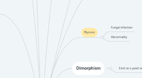
10. Dimorphism
10.1. Exist as a yeast and mold
11. Mycosis
11.1. Fungal Infection
11.2. Abnormality
12. Saprophytic
12.1. Environmental decomposers
12.1.1. Feed on dead organic matter
13. Clinical grouping
13.1. Cutaneous mycoses
13.1.1. Skin, hair, nails
13.1.2. Keratin/protein
13.1.3. Species
13.1.3.1. Epidermaphyton
13.1.3.1.1. No microconidia
13.1.3.1.2. Cluster of macroconidia
13.1.3.2. Microsporan
13.1.3.3. Trichophyton
13.1.3.4. Cause
13.1.3.4.1. Tinea (ringworms)
13.2. Idphor
13.3. Subcutaneous mycoses
13.3.1. Usually enter by trauma
13.3.2. Can be serious if not treated
13.3.3. Species
13.3.3.1. Sporothrix schenchkii
13.3.3.1.1. Disease
13.3.3.2. Bacteria like
13.3.3.2.1. Actinomyces
13.3.3.2.2. Nocardia
13.3.3.2.3. Disease
13.4. Many start with respiratory complication
13.5. Blastomycosis dermatidis
13.5.1. Blastomycosis
13.5.1.1. Found central and southeastern US
13.5.1.2. Diagnostic
13.5.1.2.1. X-ray
13.5.1.2.2. Acid fast
13.5.1.3. Inhalation of spores
13.5.1.3.1. Skin penetration
13.5.1.3.2. Air
13.5.1.4. Self-limiting
13.5.1.5. Dimorphic fungi
13.5.1.6. Morphology
13.5.1.7. Mimic TB
13.5.1.7.1. Wide base budding yeast cells
13.6. Systemic mycoses
13.6.1. Usually affect only 1% of healthy bodies
13.6.2. Affects multiple systems
13.6.3. Tissues, muscle, and bone
13.6.4. Most serious in respiratory & nervous system
13.6.5. Species
13.6.5.1. Coccidioides immitis
13.6.5.1.1. Coccidioidomycosis
13.6.5.2. Histoplasm capsulatum
13.6.5.2.1. Self-limiting
13.6.5.2.2. Histoplasmosis
13.6.5.3. Cryptococcus neoformans
13.6.5.3.1. Cryptococcosis
13.7. Classified by level of involvement in bodies
13.8. Oppotunistic mycoses
13.8.1. Does not ordinarily cause disease but if immunocompromised, disease can rise
13.8.1.1. Usually seen when patients are on antibotics
13.8.2. Aseptic hyphae
13.8.3. Species
13.8.3.1. Rapid growers
13.8.3.1.1. Rhizopus
13.8.3.1.2. Mucor
13.8.3.1.3. Absidia species
13.8.3.1.4. Causes
13.8.3.2. Candida
13.8.3.2.1. Candidiasis
13.8.3.3. Aspergillus
13.8.3.3.1. Aspergillosis
14. Cultures
14.1. Skin scrapings
14.2. Specimens
14.2.1. Hair
14.2.2. Nails
14.2.3. Sputum
14.2.4. Cerebrospinal fluid
14.2.5. Drainage from lesions
14.3. Diagnostic Tools
14.3.1. Sabouraud's agar
14.3.1.1. Selective for fungal agents
14.3.1.2. High sugar and low pH (acidic environment)
14.3.1.3. Antibiotics- inhibit bacterial growth
14.3.2. Fungal smear
14.3.2.1. KOH Prep
14.3.2.1.1. 10% Potassium hydroxide
14.3.2.2. Lactophenol cotton blue
14.3.2.2.1. Tape & dye, microscopic ID
