
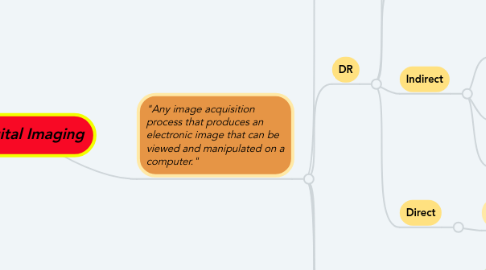
1. "Any image acquisition process that produces an electronic image that can be viewed and manipulated on a computer."
1.1. CR (PSP)
1.1.1. X-ray exposure absorbed by phosphor atoms (barium fluorohalide)
1.1.1.1. Small E loss in form of UV light
1.1.1.1.1. Other e caught in valence&conduction bands, "Stored Energy" = latent image
1.1.2. Base layer
1.1.2.1. Phosphor layer (BaFX)
1.1.2.1.1. Conductive, light blocking, protective layers
1.1.3. Cassette loaded into reader, IP removed from cassette
1.1.3.1. Solid state laser (red) scans in raster pattern
1.1.3.1.1. Light guide directs emitted light to photodetector
1.2. DR
1.2.1. Detectors initialized for exposure (rotor)
1.2.1.1. Exit energy captured for conversion to e- signal
1.2.1.1.1. Captured signal "read" before termination of exposure
1.2.2. 2-D array of DEL's (detector elements) coated on glass substrate
1.2.2.1. Pixel (x-ray/light sensitive area)
1.2.2.1.1. Fill factor = space available to capture x-ray (80% of DEL)
1.2.3. Indirect
1.2.3.1. Flat Panel Detector (Amorphous-Silicon)
1.2.3.1.1. exit beam absorbed by scintillator (CsI)
1.2.3.1.2. Scintillator (1st layer)
1.2.3.2. Charge-Coupled Device (CCD)
1.2.3.2.1. Silicon matrix
1.2.3.3. Complementary metal-oxide semiconductor (CMOS)
1.2.3.3.1. Scintillation-semiconductor
1.2.4. Direct
1.2.4.1. Flat Panel Detector (Amorphous-Selenium)
1.2.4.1.1. Photoconductor layer (a-Se) absorbs exit beam, generates proportionate e- charge
1.2.4.1.2. Photoconductor (1st layer)
1.3. Detector Response
1.3.1. Dynamic Range
1.3.1.1. "Response of detector system to varying levels of xray energy in the exit beam"
1.3.1.1.1. Linear
1.3.2. Exposure Latitude
1.3.2.1. "Range of exposures which can produce acceptable image within ALARA"
1.3.2.1.1. Tolerance for over or under exposure
1.4. Detective Quantum Efficiency (DQE)
1.4.1. "Measure of efficiency/accuracy of a detector to convert input signal (xray) to useful output data (image)
1.4.1.1. 1.0 DQE= ideal. Higher DQE= higher quality image w/ less dose
1.4.1.1.1. Image contains all data, no loss of info
1.5. Signal to Noise Ratio (SNR)
1.5.1. "Quality of signal (amount of exposure received by IR) as compared to various forms of static"
1.5.1.1. Signal=good, Noise=bad
1.5.1.1.1. Noise detracts from detail visibility
1.6. Contrast to Noise Ratio (CNR)
1.6.1. "Assessment of impact of noise on contrast resolution"
1.6.1.1. Low CNR lessens ability to distinguish between tissues which attenuate beam similarly
1.6.1.1.1. Increase SNR = increase CNR