
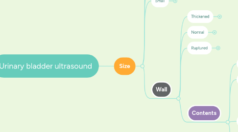
1. Size
1.1. Enlarged
1.1.1. subjective - if it fill most of the abdomen (extend cranial to umbilicus)?
1.1.1.1. Urethral obstruction
1.1.1.2. Well trained and "holding it"
1.1.1.3. Neurologic
1.2. Normal
1.2.1. highly variable
1.3. Small
1.3.1. often difficult to locate, look just cranial to pubis
1.3.1.1. forgot to clamp urinary catheter
1.3.1.2. ruptured
1.3.1.3. cystitis
1.3.1.4. just peed
1.4. Wall
1.4.1. Thickened
1.4.1.1. Diffuse
1.4.1.1.1. contains gas
1.4.1.1.2. cystitis
1.4.1.2. Multifocal
1.4.1.2.1. Often polypoid with narrow stalk and intraluminal projections. Often at the apex.
1.4.1.3. Focal
1.4.1.3.1. mural thickening, irregular mucosal margins.
1.4.2. Normal
1.4.2.1. < 4 mm, uniform thickness, no distinct layering
1.4.3. Ruptured
1.4.3.1. Discontinuous wall or can't locate the bladder at all. Often appears intact on ultrasound despite being ruptured.
1.4.4. Contents
1.4.4.1. Normal
1.4.4.1.1. anechoic urine
1.4.4.2. Non-dependent hyperechoic
1.4.4.2.1. gas
1.4.4.3. Dependent
1.4.4.3.1. well margined, hyperechoic with acoustic shadowing, can see twinkle artifact
1.4.4.3.2. hyperechoic, amorphous, swirls with motion
1.4.4.3.3. well margined or amorphous, medium echoic. Absent Doppler signal
1.4.4.4. Suspended (echogenic)
1.4.4.4.1. fat droplets
1.4.4.4.2. active sediment
1.4.4.4.3. artifact (clutter)
1.4.4.4.4. debris/casts