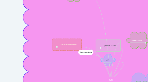
1. risk factors
1.1. sepsis, contrast media, exposure to nephrotoxins. glomerulonephritis and acute tubular necrosis (ATN)- hypovolemic (sudden drop in BP or volume),
2. medications
3. patho:
4. prerenal causes
4.1. intrarenal causes
4.1.1. postrenal causes
4.1.1.1. sudden obstruction of urine, tumor, injury, enlarged prostate. TX: find obstruction and treat obstruction. occurs cuz of bilateral obstruction of structures leaving the kidney. causes- mechanical obstruction of the lower urinary tract (ureters and bladder). flow of urine is obstructed at some point at the ureters for below = reflux into the renal pelvis, impaired kidney function. causes include: BPH, prostate cancer, calculi, trauma, and external tumors. - reversible if obstruction is removed before kidney damage if not removed AKI occurs.cause not identified = intrarenal parenchymal damage occurs. risk factors: stone, tumor, bladder atony, prostate hyperplasia, urethral stricture, spinal cord disease or injury.
4.1.2. occurs cuz of direct damage to the kidney from lack of oxygen, indicating damage to the glomeruli, nephrons, or tubules. toxins, drugs, nephrotoxic drugs, direct infections, acute tubular necrosis.- TX: figure out what is causing it treat the cause to treat the acute. risk factors: physical injury- trauma, hypoxic injury: renal artery or vein stenosis or thrombosis, chemical injury- acute nephrotoxins (antibiotics, contrast dye, heavy metals, blood transfusion reaction, alcohol, cocaine), immunologic injury- infection, vasculitis, acute glomerulonephritis.
4.2. occurs cuz of volume depletion, and prolonged reduction of blood flow to the kidneys= ischemia of nephrons. occurs before damage to the kidneys. early intervention restoring fluid volume deficit can reserve AKI and prevent CKD. result of external factors that reduce renal blood flow & lead to decreased glomerular perfusion and filtration, also affects azotemia. results in low urine, increased BUN and creatinine, inability to conserve water. if it is due to loss of volume replace fluids. risk factors: renal vascular obstruction, shock, decreased cardiac output = decreased renal perfusion, sepsis, hypovolemia, peripheral vascular resistance, use of aspirin, ibuprofen, or NSAIDs, liver failure.
5. clinical manifestations
5.1. onset: sudden (hrs-days)
5.1.1. nephron damage: 50%
5.1.2. duration:2-4 wks <3 months
5.1.3. prognosis: reversible
5.1.4. etiology: pre-renal, intrarenal, post-renal causes
5.2. findings are related to waste build up and decreases urine output. most every body system can be affected. cardio: hypertension, fluid overload (dependant & generalized edema), dysrhythmias (hyperkalemia). resp: crackles, decreased oxygenation, shortness of breath. renal: scant to normal or excessive urine output, depending on the phase; possible hematuria. neuro: lethargy, muscle twitching, seizures. integ: dry skin & mucous membranes. -assess for findings associated with underlying causes. Book: urine volume decreases (initiating & oliguric phase.), fluid volume overload- due to decreases in urine output. Increased K+- due to impaired excretion. decreased Na+- inability to conserve sodium. decreased calcium and increased phosphorus- decreased GI absorption of Ca+ due to the kidneys inability to activate vit D, decreased Ca+ stimulates the parathyroid gland to release parathyroid hormone = stimulates the release of Ca+ and phosphorus from bone, decreased phosphorus clearance. Metabolic acidosis: decreased acid excretion, increased loss of of Bicarbonate, increased production of nonvolatile acids such as lactic acid or phosphoric acid. Anemia: impaired erythropoietin production. Increased BUN: impaired clearance, dehydration, increased protein intake and breakdown. Increased creatinine: impaired clearance best indicator of renal failure.
6. management
6.1. lab tests: electrolytes: Na+ decreased (prerenal azotemia) or increased (intrarenal azotemia), hyperkalemia, hyperphosphatemia, hypocalcemia; urine specific gravity varies in postrenal type; can be elevated up to 1.030, in prerenal or diluted as low as 1.000 in intrarenal. hematocrit-decreased, UA- presence of sediment (RBC, casts), ABG: metabolic acidosis.BUN can increase to 8-100 within 1 wk. creatinine levels increase 1-2 every 24-48 hrs or 1-6 in 1 wk or less
6.2. diagnostic tests
6.3. kidney biopsy: done when the cause of AKI is unknown, also to detect immunological diseases, or determine kidney dysfunction reversibility and need for dialysis. imaging: X-ray- (KUB) to detect calculi and hydronephrosis & to determine size of kidneys. Ultrasound: to detect and obstruction in urinary tract. CT scan: w/o contrast dye or MRI to detect anatomical changes, tumors, or other obstructions, patency of ureters; renal perfusion. Nuclear meds: cystography, retrograde pyelography.
7. nursing assessment /intervention assessment
8. nursing interventions assessments/actions
9. diagnostic tests
10. phases of AKI
10.1. initiating/onset phase
10.1.1. oliguric phase
10.1.1.1. diuretic phase
10.1.1.1.1. recovery phase
10.1.1.1.2. when healing begins,begins when the kidneys start to recover; diuresis of a large amount of fluid occurs; can last for 2-6 wks
10.1.1.2. when urine output decreases because of renal tissue damage. begins w/ kidney insult; urine output is 11-400 ml/24 hr w; W/O diuretics; and lasts for 1-3 wks
10.1.2. when kidney injury first occurs, begins with the onset of the even, ends when oliguria develops and lasts for hours to days.
