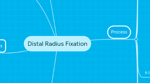
1. Place
1.1. The Johns Hopkins Medical Insititute
1.2. Zayed Building OR 303
2. People
2.1. Attending
2.1.1. Dr. Hasenboehler
2.2. Fellow
2.3. Resident
2.4. Circulating Nurse
2.5. Scrub Nurse
2.6. Anesthesiologist
2.7. C-Arm Technician
2.7.1. Charles
2.8. Orthopaedic company representative
2.9. Patient
2.9.1. Has had previous fractures of the distal radius
2.9.2. losing degrees of freedom after 2 fractures
3. Products
3.1. Standard Anesthesiology Equipment
3.2. Patient Bed
3.2.1. Lateral arm addition
3.3. Surgical Tool Table
3.4. Implant Equipment
3.4.1. Implants
3.4.1.1. Plates
3.4.1.2. Screws
3.4.2. Drills
3.4.3. Guide Wires
3.5. C-arm
3.6. Cautery Tools
3.7. Suctioning Equipment
4. Basic Process
4.1. Patient Anesthetized
4.2. Incision made above distal radius
4.3. Clearing of callus formed around break
4.4. Plate inserted across fracture
4.5. Two screws inserted
4.6. Position confirmed
4.7. Remaining screws placed
4.8. Screw Placement and Plate location confirmed
4.9. Suturing to close incision
4.10. Patient woken up and moved to PACU
5. Process
5.1. 7:50 am
5.1.1. Patient enters room on bed
5.1.1.1. anesthesia started
5.1.2. Nurse maneuvers bed next to surgical bed
5.1.2.1. Nurse attempts to lock bed in place
5.1.2.1.1. Takes 3 attempts before brake is secured
5.1.2.1.2. Brake is located on wheels
5.1.2.1.3. Intended for foot to be used to activate locking mechanism
5.1.2.1.4. Foot pedal is very small and hard to locate blind
5.1.2.1.5. Foot pedal is awkward to push down
5.1.3. Nurse, Resident, Fellow, and circulating nurse move patient to surgical bed
5.1.3.1. two people on either side of beds
5.1.3.2. two leaning over surgical bed to pull patient towards them, two patients lift patient towards surgical bed
5.1.3.3. No on scrubbed but with gloves on
5.1.4. Nurse adds appendage attachment to surgical table
5.1.4.1. Nurse carries in table attachment
5.1.4.1.1. its heavy and bulky
5.1.4.2. attachement slides onto fixation device on side of table
5.1.4.3. tightened in place blindly
5.1.4.3.1. table inhibits seeing attachment below it
5.1.4.4. attachement is bouncy
5.1.4.4.1. single stabilizing leg with spring loaded foot
5.2. 8:04 am
5.2.1. While Patient is prepped and table is adjusted, Doctors scrub up
5.2.1.1. doctors enter room
5.2.1.2. gown is presented by scrub nurse
5.2.1.2.1. doctor inserts arms into sleeves and pulls gown on
5.2.1.3. two sets of gloves are put on by doctors
5.2.1.3.1. aids in preventing BBP transmission in the event of cuts
5.2.1.4. two separate ties
5.2.1.4.1. one with two internal ties on the backside of sterile gown
5.2.1.4.2. another attached to tab which nurse holds while doctor spins
5.3. 8:17 am
5.3.1. Patients two fingers are wrapped in cloth strips and hung from IV stand
5.3.1.1. allows arm to be suspended in order to sterilize arm
5.3.1.2. IV stand is not stable
5.3.2. Iodine pen cracked to activate
5.3.2.1. Iodine rubbed all over patients arm
5.3.2.2. Is this comprehensive?
5.3.3. tourniquet applied to arm to prevent excessive bleeding
5.3.4. Sterile drapes places all over patients body
5.3.4.1. drape with elastic hole stretch and pulled up patients arm
5.4. 8:19 am
5.4.1. Table attachement issues
5.4.1.1. balance leg is now off the ground
5.4.1.1.1. does not provide stable surgical surface
5.4.2. surgical tool table moved closer to patient
5.5. 8:31 am
5.5.1. scrub hands fellow scalpel
5.5.2. fellow makes first incision
5.5.2.1. cuts through skin
5.5.2.2. cuts through fascia
5.5.2.3. cuts through muscle
5.5.3. cautery tool used to stop bleeders
5.5.3.1. minimal bleeding
5.5.3.2. done by touching cauterizer to tweezers
5.5.3.2.1. easier to grip bleeders
5.5.4. some suction used
5.6. 8:37 am
5.6.1. Mini retractors placed on tissue to expose deep tissue
5.6.2. Synthes screw driver used to place k-wire into thumb
5.6.2.1. white piece of unknown substance fell out of drill
5.6.2.1.1. possible contamination?
5.6.3. Second k-wire drilled into radius proximal to fracture
5.6.4. distractor structure slides over two k-wires and fixed into place
5.6.4.1. used to apply decompressive or compressive forces across fracture
5.6.4.1.1. adds additional injuries beyond main surgical site
5.6.4.1.2. can it be done without it?
5.6.5. x-ray with c-arm taken to visual fracture location
5.6.5.1. about 10 taken
5.6.5.2. needs to be reduce to decrease radiation exposure
5.6.6. Callus is slowly chipped away using kerrison punch and ronjuers
5.6.6.1. as pressure is applied to wrist, the hand moves
5.6.6.2. need a second person to hold wrist and hand down
5.6.6.3. could cause accidental injury to patient or ORP
5.7. 8:57 am
5.7.1. Discuss with Dr. Hasenboehler about patient's history while callus is being removed
5.8. 9:03 am
5.8.1. using chisel and mallet to chip away callus
5.8.1.1. wrist is bouncing back with every mallet hit
5.8.1.2. decrease force actually applied to callus
5.8.1.2.1. force displaced to table foot spring
5.8.2. use pick and mallet to chip away callus
5.8.2.1. k-wire not supposed to be used for mallet and chipping away of bone
5.8.2.2. can't get it out
5.8.2.2.1. must use plier to rip k-wire out
5.9. 9:24 am
5.9.1. C-arm takes x-ray
5.9.1.1. articular surfaces do not line up
5.9.1.1.1. every time surfaces do not line up, more x-rays
5.9.2. two k-wires drilled directly into bone on either side of fracture
5.9.2.1. x-ray to verify position
5.9.3. plate is slid over k-wires
5.9.3.1. k-wires removed
5.9.4. first screw is drilled into most proximal end of plate
5.9.5. second screw is drilled into most distal end of plate
5.9.5.1. slightly less angled than guide so as to not interfere with joint space
5.9.5.1.1. can lead to complications, done so as to not interfere with joint
5.10. 9:44 am
5.10.1. while screws are being placed, discussed patient history with Dr. Hasenboehler
5.10.1.1. originally treated conservatively
5.10.1.2. resulted in dorsal migration
5.10.1.2.1. loss of degrees of freedom
5.10.1.3. plate is being used to prevent that
5.11. 9:49 am
5.11.1. x-rays taken to verify screw placement
5.11.1.1. slightly convergent
5.11.1.1.1. need to be redone for strength purposes
5.11.1.1.2. screws will be used, simply repositioned
5.11.2. screw placement is guess and check
5.11.2.1. must avoid joint space
5.11.2.2. checked iteratively with x-ray until correct alignment achieved
5.11.3. the rest of screws are placed one after another
5.11.4. screws checked with x-ray
5.11.4.1. one is suspiciously close to joint space
5.11.4.2. screw is removed and replaced with a more conservative angle
5.11.5. tourniquet is released
5.11.5.1. bleeders cauterized
5.11.5.2. suctioning of fluids
5.11.5.3. arm swells
5.11.5.3.1. reason sutures are not done before because sutures would result into too much tension on tissue
5.12. 10:03 am
5.12.1. "distractor hits unpredictable veins" - Hasenboehler
5.12.2. "no good way to suture muscle" - Hasenboehler
5.12.3. Begin suturing deep tissues and fascia with resorbable sutures
5.12.4. suture skin
5.12.4.1. often staples are used as well
