1. Congenital malformations (by gross and micro appearances)
1.1. MOST COMMON
1.1.1. Pancreas divisum
1.1.1.1. failure of fusion
1.1.1.1.1. Dorsal and ventral duct systems
1.2. Other Congenital Pancreatic Anomalies
1.2.1. Congenital pancreatic cysts
1.2.1.1. Often multiple
1.2.1.2. almost always a/w underlying congenital disease
1.2.1.2.1. Von Hippel-Lindau syndrome
1.2.2. Ectopic Pancreas
1.2.2.1. Where?
1.2.2.1.1. Stomach and duodenum (MC)
1.2.2.1.2. jejunum, meckel diverticula, ileum
1.2.2.2. What?
1.2.2.2.1. Aberrantly situated pancreatic tissue
1.2.3. Annular pancreas
1.2.3.1. ring of normal pancreas completely encircling second portion of duodenum
1.2.3.1.1. May cause obstruction
2. Acute vs chronic pancreatitis
2.1. gross and macroscopic appearance
2.2. underlying causes and pathogenesis
2.3. genetic predisposition
2.4. morphology
2.4.1. inflammation
2.4.2. fat necrosis
2.4.3. parenchymal destruction
2.4.4. hemorrhage
2.5. clinical signs and symptoms
2.6. complications
2.7. outcomes
2.8. Pancreatitis
2.8.1. Acute Pancreatitis
2.8.1.1. Main Causes 80%
2.8.1.1.1. Males
2.8.1.1.2. Females
2.8.1.2. Morphology
2.8.1.2.1. Microscopic
2.8.1.2.2. Gross
2.8.1.3. Clinical Features
2.8.1.3.1. Plasma findings
2.8.1.3.2. Signs / Symptoms
2.8.1.3.3. Diagnosis of Acute Pancreatitis
2.8.1.4. Complications
2.8.1.4.1. ARDS
2.8.1.4.2. ARF
2.8.1.4.3. Pancreatic Abscess
2.8.1.4.4. Pancreatic pseudocyst
2.8.1.5. Treatment
2.8.1.5.1. Supportive
2.8.1.5.2. No PO intake
2.8.1.6. Pathophysiology of Pancreatitis
2.8.1.6.1. hemorrhage
2.8.1.6.2. Necrosis
2.8.1.6.3. parenchymal destruction
2.8.1.6.4. Inflammation
2.8.2. Chronic pancreatitis
2.8.2.1. Causes / Risk Factors
2.8.2.1.1. Other Risk Factors
2.8.2.1.2. Causes
2.8.2.2. Gross appearance
2.8.2.2.1. fibrosis
2.8.2.2.2. atrophy
2.8.2.2.3. dilated ducts with calcified concretions
2.8.2.3. Imaging
2.8.2.3.1. "Chain of Lakes"
2.8.2.4. Microscopic appearance
2.8.2.4.1. fibrosis
2.8.2.4.2. atrophy
2.8.2.4.3. chronic inflammation
2.8.2.5. Outcomes
2.8.2.5.1. Atrophy
2.8.2.5.2. Chronic pain
2.8.2.5.3. Pancreatic cancer
2.8.2.6. Lab Findings
2.8.2.6.1. Tests only detect advanced disease
2.8.3. Autoimmune pancreatitis
2.8.3.1. Associated with
2.8.3.1.1. IgG4-secreting plasma cells in pancreas
2.8.3.2. Signs and Symptoms
2.8.3.2.1. mimic pancreatic cancer
2.8.3.2.2. i.e. obstructive jaundice
2.8.3.3. Responds to steroids
2.8.3.4. Histology
2.8.3.4.1. periductal inflammation
2.8.3.4.2. periductal fibrosis
2.8.3.4.3. periductal narrowing
2.8.4. Pathogenesis
2.8.4.1. Normal
2.8.4.1.1. Zymogens synthesized
2.8.4.1.2. Trypsin activates proenzymes
2.8.4.1.3. Trypsin inhibitors
3. Pancreatic neoplasms
3.1. gross and histologic appearance
3.2. location
3.3. risk factors
3.4. clinical findings
3.5. Side effects
3.6. Genetic mutations
3.7. outcomes / survival rates
3.8. Pancreatic cystic neoplasms
3.8.1. Serous
3.8.1.1. Serous cystadenomas
3.8.1.1.1. F > M
3.8.1.1.2. Microcystic
3.8.1.1.3. almost always benign
3.8.2. Mucinous
3.8.2.1. Mucinous cystic neoplasm
3.8.2.1.1. F - almost exclusively
3.8.2.1.2. Macrocystic
3.8.2.1.3. Location
3.8.2.1.4. Benign or malignant
3.8.3. Intraductal Papillary Mucinous
3.8.3.1. IPMN
3.8.3.1.1. M > F
3.8.3.1.2. Location
3.8.3.1.3. Mucin accumulation leads to cystic dilatation
3.8.3.1.4. Intraductal proliferation of mucinous cells in papillary arrangements
3.9. Pancreatic ductal adenocarcinoma
3.9.1. Epidemiology
3.9.1.1. AfAm > White
3.9.1.2. M > F
3.9.1.3. 4th leading cause of cancer death in U.S.
3.9.1.3.1. 5-year survival rate
3.9.2. Risk Factors
3.9.2.1. MAIN
3.9.2.1.1. Smoking
3.9.2.2. Chronic pancreatitis, DM
3.9.2.3. Diets high in fat / protein
3.9.2.4. Alcohol
3.9.2.5. Genetic mutations (inherited and acquired)
3.9.2.5.1. BRCA2 mutation
3.9.2.5.2. Lynch Syndrome - HNPCC
3.9.2.5.3. KRAS mutation - 90-95%
3.9.2.5.4. CDKN2A mutation - 95%
3.9.3. Progression
3.9.3.1. Preceded by
3.9.3.1.1. PanIN
3.9.3.2. progression from normal to PanIN to invasive tumor
3.9.3.3. Morphological progression coincides with Genetic progression
3.9.3.3.1. KRAS and CDKN2A mutations
3.9.4. Morphology
3.9.4.1. Location
3.9.4.1.1. 60% - Head
3.9.4.1.2. 20% - Diffuse
3.9.4.1.3. 15% - Body
3.9.4.1.4. 5% - Tail
3.9.4.2. Microscopic
3.9.4.2.1. high degree of invasiveness
3.9.4.2.2. desmoplastic
3.9.4.2.3. Malignant glands, arising from ductal epithelium of pancreas with abundant desmoplasia
3.9.5. Clinical Findings
3.9.5.1. Commonly unresectable
3.9.5.2. Signs and Symptoms
3.9.5.2.1. Pain
3.9.5.2.2. Weight Loss
3.9.5.2.3. Obstructive jaundice
3.9.5.2.4. Migratory Thrombophlebitis
3.9.5.2.5. Labs
3.9.5.2.6. Courvoisier's Sign
3.10. Pancreatic endocrine neoplasms
3.10.1. Insulinoma (MOST COMMON PEN)
3.10.1.1. Beta-Cell Tumors
3.10.1.1.1. Severe hypoglycemia
3.10.1.1.2. Whipple Triad
3.10.1.1.3. Small and encapsulated
3.10.1.1.4. 10% metastasize
3.10.2. Gastrinoma
3.10.2.1. G-Cell Tumors
3.10.2.1.1. Location - Gastrinoma Triangle
3.10.2.1.2. Zollinger-Ellison syndrome
3.10.2.1.3. over 50% are locally invasive or metastasized at time of diagnosis
3.10.3. Glucagonoma
3.10.3.1. Alpha-Cell Tumors
3.10.3.1.1. Glucagonoma syndrome
3.10.3.1.2. 50% have metastases at time of diagnosis
3.10.3.1.3. F
3.10.4. May be associated with
3.10.4.1. MEN 1 syndrome
3.10.4.1.1. Parathyroid
3.10.4.1.2. Pancreas (islet cells)
3.10.4.1.3. Pituitary
3.10.5. Morphology
3.10.5.1. Gross
3.10.5.1.1. well-circumscribed
3.10.5.1.2. yellow tumor
3.10.5.1.3. Tail of pancreas
3.10.5.2. Microscopic
3.10.5.2.1. Uniform, monotonous tumor cells
3.10.5.2.2. Forming nests and trabeculae
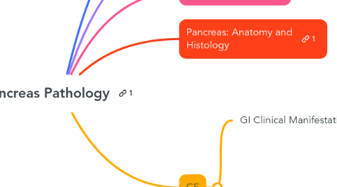
4. Pancreas: Anatomy and Histology
5. CF
5.1. GI Clinical Manifestations
5.1.1. Intestinal obstruction
5.1.2. Pancreatic fibrosis and insufficiency
5.1.2.1. malabsorption
5.1.3. Biliary cirrhosis
5.2. Treatment
5.2.1. PERT
5.2.1.1. Pancreatic Enzyme Replacement Therapy
5.2.1.2. Mechanism
5.2.1.2.1. Contains enzymes (lipase, protease, amylase) to help ensure nutrients are effectively absorbed from food
5.2.1.2.2. coating dissolves in alkaline medium of duodenum
