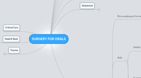
1. Critical Care
2. Head & Neck
2.1. Nodule
2.1.1. Midline
2.1.1.1. Thyroglossal Duct Cyst
2.1.1.1.1. US & FNA
2.1.1.1.2. Moves w tongue protrusion
2.1.1.2. Thyroid Nodule
2.1.1.2.1. US
2.1.1.2.2. TFTs
2.1.1.2.3. FNA
2.1.1.2.4. FHx or H/o Radiation
2.1.1.2.5. <4cm
2.1.2. H&P
2.1.2.1. H/o neck RTx
2.1.2.2. Fam Hx
2.1.2.2.1. Endocrinopathy
2.1.2.2.2. Cancers
2.1.2.3. LNs
2.1.2.4. Risk factors
2.1.2.4.1. Tobacco
2.1.2.4.2. EtOH
2.1.3. Work-Up
2.1.3.1. TSH/ TFTs/ Ca
2.1.3.2. FNA
2.1.3.2.1. US guided
2.1.3.2.2. Abnormal LN also
2.1.3.2.3. Indeterminate or NonDx
2.1.3.3. Voice Changes
2.1.3.3.1. Laryngoscopy
2.1.4. Parotid mass
2.1.4.1. Imaging
2.1.4.2. No FNA
2.1.4.2.1. Superficial Parotidectomy
2.2. Hyperthyroidism
2.2.1. Grave's Dz
2.2.1.1. ABs to TSHR
2.2.1.1.1. LATS and TSIs
2.2.1.2. Medical Tx
2.2.1.2.1. PTU
2.2.1.2.2. Methamizol
2.2.1.2.3. Propranolol
2.2.1.3. Surgery If
2.2.1.3.1. Recurrent
2.2.1.3.2. Severe Hyperthyroid
2.2.1.3.3. Age>55
2.2.1.3.4. Symptomatic goiter
2.2.1.3.5. Or Radioablation
2.2.2. Multinodular Goiter
2.2.2.1. Surgery vs. RIA
2.2.2.1.1. Surgery if compressive Sx
2.2.2.1.2. Awake fiberoptic intubation
2.2.2.2. Euthyroid
2.2.2.2.1. RIA works 50%
2.2.3. Toxic Adenoma
2.2.3.1. Hot on T-Scan
2.2.3.1.1. Resect
2.2.3.1.2. RIA if warranted
2.2.4. Thyroid Storm
2.2.4.1. Sx
2.2.4.1.1. Hyperthermia
2.2.4.1.2. MS Changes
2.2.4.1.3. Diarrhea, N/V
2.2.4.1.4. Tachycardia
2.2.4.2. Tx
2.2.4.2.1. Propranolol
2.2.4.2.2. PTU
2.2.4.2.3. Dexamethasone
2.3. Hyperparathyroidism
2.3.1. Primary
2.3.1.1. Adenoma
2.3.1.2. Hyperplasia
2.3.2. Secondary
2.3.3. Tertiary
3. Trauma
3.1. Liver
3.1.1. Blunt
3.1.1.1. Grade I-II
3.1.1.1.1. Serial H/H
3.1.1.2. Grade III-IV
3.1.1.2.1. ICU Admit
3.1.1.3. +Blush on CT
3.1.1.3.1. Angio-Embolization
3.2. Rectum
3.2.1. Procto
3.2.2. Laparotomy
3.2.2.1. Extraperitoneal Inj
3.2.2.1.1. Primary Repair if Feasible
3.2.2.1.2. Loop Colostomy
3.2.2.1.3. Presacral Drainage
3.2.2.2. Intraperitoneal Inj
3.2.2.2.1. Repair
3.2.2.2.2. Resection& Anastomosis
3.2.2.3. GU Involved
3.2.2.3.1. Repair both
3.2.2.3.2. Divert Both
3.2.2.3.3. Interpose tissue
3.2.2.4. Anal Sphincter Involved
3.2.2.4.1. Sphincteroplasty after healing
3.3. Thoracic
3.3.1. Transmediastinal GSW
3.3.1.1. HD unstable
3.3.1.1.1. To OR
3.3.1.1.2. No pericardiocentesis
3.3.1.2. Post-Repair
3.3.1.2.1. Esophagram
3.3.2. Subclavian inj
3.3.2.1. Unclear
3.3.2.1.1. Median sternotomy
3.3.2.2. Left
3.3.2.2.1. Unstable
3.3.2.2.2. Stable
3.3.3. Tracheal Injury
3.3.3.1. Cervical
3.3.3.1.1. Collar incision
3.3.3.2. Thoracic
3.3.3.2.1. R Thoracotomy
3.4. Pelvic Fx
3.4.1. Stable
3.4.2. Unstable
3.4.2.1. Absent Intraabdominal Issues
3.4.2.1.1. Pre-Peritoneal Pelvic Packing
3.4.2.1.2. Angio if available
3.4.2.2. FAST+ or possible peritonitis
3.4.2.2.1. Ex-Lap
3.5. Diaphragmatic Inj
3.5.1. Primary repair
3.5.1.1. Non-absorb sutures
3.5.1.2. #o
3.5.1.3. Interrupted horizontal mattress
3.5.2. Large defect
3.5.2.1. Goretex Graft
3.5.2.2. Porcine Graft
3.5.2.2.1. If contaminated
4. Genitourinary
4.1. Kidney
4.2. Ureter
4.2.1. Injury
4.2.1.1. Proximal
4.2.1.1.1. Ureteroureterostomy
4.2.1.1.2. Ureteropelicostomy
4.2.1.1.3. Ureterocalicostomy
4.2.1.2. Distal
4.2.1.2.1. Ureter does not reach
4.3. Bladder
4.4. Gonads
4.4.1. Testes
4.4.2. Ovaries
4.5. Pudenda
4.5.1. Penis
4.5.2. Vagina
5. Breast
5.1. Mass
5.1.1. Fibroadenoma
5.1.1.1. Core Bx
5.1.1.1.1. Excise if
5.1.2. Cyst
5.1.2.1. Simple
5.1.2.1.1. Aspiration
5.1.2.2. Complex
5.1.2.2.1. US guided Biopsy
5.2. Pain
5.3. Nipple discharge
5.3.1. PE focus on culprit quadrant
5.3.2. Calculate GAIL Risk
5.3.3. Imaging
5.3.4. Fluid type
5.3.4.1. Serous/bloody
5.3.4.1.1. If duct ID'd
5.3.4.1.2. If not
5.3.4.2. Milky
5.3.4.2.1. Prolactin level
5.3.4.3. Do not send it for Histology
5.3.4.4. No galactogram
5.3.5. Concerning
5.3.5.1. Spontaneous
5.3.5.2. Unilateral
5.3.5.3. Recurrent
5.4. Screening
5.4.1. Self exam @20 Qm
5.4.1.1. Clinical exam @3y
5.4.1.2. Start @40
5.4.1.2.1. MMG & CE yearly
5.4.2. MRI if
5.4.2.1. BRCA +
5.4.2.2. Li-Fraumeni
5.4.2.3. Radiation to chest
5.4.2.4. Calculated lifetime risk >25%
5.5. Infection
5.5.1. PO ABx
5.5.2. I&D if abscess
5.5.2.1. Excise/Bx if recurrent
5.5.3. Lactation
6. Thoracic
6.1. Esophagus
6.1.1. Motility Disorders
6.1.1.1. Esophagram
6.1.1.2. PH Probe
6.1.1.3. EGD
6.1.1.4. Manometry
6.1.1.5. TREATMENT
6.1.1.5.1. DES
6.1.1.5.2. Nutcracker Eo
6.1.2. Diverticuli
6.1.2.1. Zenker's
6.1.2.1.1. Endoscopic
6.1.2.1.2. Open Resection
6.1.2.1.3. EGD
6.1.2.2. Parabronchial Diverticulum
6.1.2.2.1. Traction
6.1.2.2.2. TB or Lung CA
6.1.3. Achalasia
6.1.3.1. Characteristics
6.1.3.1.1. Medical
6.1.3.1.2. Endoscopic
6.1.3.1.3. Surgical
6.1.4. Stricture
6.1.4.1. R/o CA
6.1.4.1.1. Bx
6.1.4.1.2. EUS
6.1.4.2. R/o Achalasia
6.1.4.2.1. Manometry
6.1.4.3. Balloon dilation
6.1.4.3.1. Nissen for GERD control
6.1.5. Barrett's
6.1.5.1. Nondisplastic
6.1.5.1.1. GERD control
6.1.5.2. LGD
6.1.5.2.1. ReBx in 3 mos
6.1.5.2.2. Treat GERD
6.1.5.3. HGD
6.1.5.3.1. Surveillance Q 3 mos
6.1.5.3.2. CA W/U
6.1.5.4. Indeterminate GD
6.1.5.4.1. Place on high dose PPI
6.1.5.5. Length
6.1.5.5.1. >3cm
6.1.5.5.2. <3cm
6.1.6. Reflux
6.1.6.1. Try Non-Surgical
6.1.6.1.1. Per-op W/U
6.1.6.1.2. Trans-Thoracic
6.1.6.1.3. Trans-abdominal
6.1.6.1.4. Short-Esophagus
6.1.7. Perforation
6.1.7.1. General Approach
6.1.7.1.1. Find it
6.1.7.1.2. Thin barium swallow
6.1.7.2. Highly unstable pt
6.1.7.2.1. Cervical esophagostomy
6.1.7.2.2. T tube drainage
6.1.8. Malignancy
6.1.9. Caustic Inj
6.1.9.1. R/o perf
6.1.9.1.1. + Perf
6.1.9.2. EGD
6.1.9.2.1. 1st degree
6.1.9.2.2. 2-3rd degree
6.2. Aorta
6.3. Cardiac
6.4. Lung
6.5. Mediastinal
7. Vascular
7.1. Peripheral arterial
7.1.1. Claudication
7.1.1.1. Exercise regimen
7.1.1.2. Quit smoking
7.1.1.2.1. Do not operate if still smoking
7.1.1.2.2. Life-limiting ischemia
7.1.2. Critical limb ischemia
7.1.2.1. Ulcer/ open wound
7.1.2.2. Rest pain
7.1.2.3. Neuropathy
7.1.3. Popliteal aneurysm
7.1.3.1. Check for other aneurysms
7.1.3.2. Asymptomatic
7.1.3.2.1. Thrombosed
7.1.3.2.2. Non-thrombosed
7.1.3.3. Symptomatic
7.1.3.3.1. Elective bypass
7.1.3.4. Limb-threatening
7.1.3.4.1. Emergent bypass
7.1.3.4.2. Fogarty thrombectomy
7.2. Peripheral venous
7.3. Carotid
7.3.1. H&P
7.3.1.1. Sx
7.3.1.1.1. Intervene if > 60%
7.3.1.1.2. Plaque ulcer
7.3.1.2. A/Sx
7.3.1.2.1. Intervene if > 70%
7.4. Abdominal aorta
7.4.1. AAA
7.4.1.1. H&P
7.4.1.1.1. C-Scope
7.4.1.1.2. Check for pop aneurysm
7.4.1.2. CT Angio
7.4.1.2.1. Offer EVAR
7.4.1.2.2. Open repair
7.4.1.3. Ruptured
7.4.1.3.1. CTA
7.4.1.3.2. Prep & Drape before induction
7.4.1.3.3. Get supra celiac control
7.4.1.4. Incidental
7.4.1.5. Post-op
7.4.1.5.1. Colonic ischemia
7.5. Acute Mesenteric Ischemia
7.5.1. CT Scan w IV
7.5.1.1. Embolic Dz
7.5.1.1.1. Suspected source
7.5.1.1.2. Evidence of end-organ injury
7.5.1.2. Thrombotic Dz
7.5.1.2.1. Source
7.5.1.2.2. Bypass to supraceliac aorta preferred
7.5.1.2.3. Bypass to inferior aorta may kink
7.6. Aorto-enteric Fistula
7.6.1. HD stable
7.6.2. Unstable
7.6.2.1. Lap
7.6.2.1.1. Prox-Dist Control
7.6.3. Any GIBleed after Aortic bypass
7.6.3.1. AEF UPO
7.7. Infected Graft
7.7.1. Extra-anatomic bypass
7.7.1.1. Then Resection
7.8. Chronic Mesenteric Ischemia
7.8.1. CT Angio
7.8.1.1. Bypass
7.8.1.1.1. Supraceliac
7.8.1.1.2. Retrograde
7.8.1.2. Endovascular stenting
8. Hernias
8.1. Inguinal
8.2. Ventral
8.3. Diaphragmatic
8.4. Laparoscopy
8.4.1. Inguinal
8.4.2. Ventral
8.4.3. Diaphragmatic
9. Hepatobiliary
9.1. Gallbladder
9.1.1. Gallstone ileus
9.1.1.1. Small laparotomy
9.1.1.1.1. Milk bowel for stone
9.1.1.1.2. Run entire bowel
9.1.1.1.3. Enterotomy to extract
9.1.1.1.4. Don't touch the GB
9.1.2. Acalculous Cholecystitis
9.1.2.1. Stable
9.1.2.1.1. Lap Chole
9.1.2.2. Unstable
9.1.2.2.1. CT or US-guided Perc Cholecystotstomy
9.1.2.2.2. IR unavailable
9.1.3. Cholecystitis ina Cirrhotic
9.1.3.1. Partial cholecystectomy
9.2. Biliary tract
9.2.1. Cholangitis
9.2.1.1. Stabilize
9.2.1.1.1. IV Cipro/Flagyl
9.2.1.2. ERCP or PTC
9.2.1.3. CBDE
9.2.1.3.1. Extract stone
9.2.1.3.2. T-Tube if pt unstable
9.2.1.4. Duodenotomy
9.2.1.4.1. Sphincterotomy
9.2.2. Primary sclerosing cholangitis
9.2.2.1. Ursodeoxycholic acid
9.2.2.1.1. Sx relief
9.2.2.1.2. Surgery
9.2.2.1.3. Transplant
9.2.3. Post-Lap Chole Jaundice
9.2.3.1. Labs
9.2.3.2. Occlusion
9.2.3.2.1. ERCP
9.2.3.2.2. PTC
9.2.3.2.3. RUQ Exploration
9.2.3.3. Stenosis
9.2.3.3.1. ERCP
9.2.3.3.2. Balloon dilation
9.2.3.4. Late presentation
9.2.3.4.1. Retained/Primary stones
9.2.4. Obstruction
9.2.4.1. Malignant
9.2.4.1.1. Unresectable Dz
9.2.4.1.2. Cannot dissect out CBD
9.2.4.2. Stone Dz
9.2.4.2.1. Choledochotomy
9.2.4.2.2. Transduodenal Sphincteroplasty
9.2.4.2.3. Unextractable stone
9.2.4.3. Post-Op Stricture
9.2.4.3.1. Choledochoplasty
9.3. Liver
9.3.1. Abscess
9.3.1.1. IV ABx
9.3.1.1.1. Colonic Source
9.3.1.1.2. H/o Cholangitis
9.3.1.2. Perc Drain
9.3.1.2.1. Not if Multiple
9.3.1.3. If peritoneal source
9.3.1.3.1. Lap for source
9.3.2. Amebic Abscess
9.3.2.1. IV Flagyl
9.3.2.1.1. No Drain/No OP
9.3.3. Variceal bleeding
9.3.3.1. Tx
9.3.3.1.1. IV octreotide
9.3.3.1.2. Vasopressin
9.3.3.1.3. EGD Banding
9.3.3.1.4. 10% failure
9.3.4. Ascites
9.3.4.1. Max med Tx
9.3.4.1.1. Lasix 160mg/day
9.3.4.1.2. Spironolactone 400mg/day
9.3.5. Cysts
9.3.5.1. Simple
9.3.5.1.1. Watch
9.3.5.2. Imaging
9.3.5.2.1. Ecchinococal
9.3.5.2.2. Congenital
9.3.5.2.3. Cystadenoma
9.3.5.3. Symptomatic
9.3.5.3.1. Perc drain
9.3.5.4. Eccinococcal
9.3.5.4.1. Albendazole
9.3.5.4.2. Open cystectomy
9.3.5.4.3. PAIR
9.3.6. Scoring Systems
9.3.6.1. Child's-Pugh
9.3.6.1.1. Components
9.3.6.1.2. Mortality
9.3.6.2. MELD
9.3.6.2.1. Components
9.3.6.2.2. 3 Mo. Mortality
9.4. Pancreas
9.4.1. Pancreatitis
9.4.1.1. Mild
9.4.1.2. Severe
9.4.1.2.1. ICU Admission
9.4.1.2.2. Blood Cx
9.4.1.2.3. Serial CT, Hb/Hct
9.4.1.2.4. Necrosis on CT
9.4.1.3. Infected
9.4.1.3.1. IV Primaxin
9.4.1.4. Hemorrhagic
9.4.1.4.1. Angio-Embo
9.4.1.5. Sequelae
9.4.1.5.1. Pseudocyst
9.4.1.5.2. Necrosis
10. Small Bowel & Colon
10.1. Oncology
10.2. Infectious
10.2.1. C. Diff Colitis
10.2.1.1. Toxic Megacolon
10.2.1.1.1. ICU Admit
10.2.1.1.2. D/C offending agent
10.2.1.1.3. IV Flagyl, PO Vanc
10.2.1.1.4. MTx Failure
10.2.1.1.5. HD Instability
10.2.1.2. Diarrhea
10.2.1.2.1. PO or IV Flagyl
10.2.1.2.2. PO Vanc
10.2.1.2.3. PR Vanc
10.2.1.2.4. PO Rifaximin
10.2.1.2.5. Fecal transplant
10.3. Diverticulitis
10.3.1. Uncomplicated
10.3.1.1. Cipro flagyl
10.3.1.1.1. Add Amp
10.3.1.2. NPO IVF
10.3.1.2.1. Until better cliniclly
10.3.2. Complicated
10.3.2.1. Abscess
10.3.2.1.1. Hinchey I
10.3.2.1.2. Hinchey II
10.3.2.1.3. Hinchey III
10.3.2.1.4. Hinchey IV
10.3.2.2. Perforation
10.3.2.2.1. Hartmann
10.3.2.2.2. Resection impossible
10.3.2.3. Fistula
10.3.2.3.1. Hartmann
10.3.2.4. Stricture
10.3.2.4.1. Do not plasty
10.3.2.4.2. New node
10.4. Diverticula
10.4.1. Duodenal
10.4.1.1. Resect if Sx
10.4.1.1.1. Stent duct if necessary to avoid it
10.4.1.1.2. Transduodenal is better
10.4.2. Meckel's
10.4.2.1. Resect only if Sx
10.4.2.1.1. Appendectomy
10.5. Bleeding
10.5.1. Approach
10.5.1.1. NGT Aspiration
10.5.1.2. Foley, IVs, Type and Cross
10.5.1.2.1. IVF Challenge 2L
10.5.1.2.2. Transfuse 2U PRBCs
10.5.1.3. DRE, Proctoscopy
10.5.1.4. Work-up
10.5.1.4.1. Nuclear Scintigraphy
10.5.1.4.2. Colonoscopy
10.5.1.4.3. Angiography
10.5.2. Diverticular
10.5.2.1. Numbers
10.5.2.1.1. 25% lifetime risk
10.5.2.2. Work-Up
10.5.2.2.1. Colonoscopy
10.5.2.2.2. Nuclear scintigraphy
10.5.2.2.3. Angio
10.5.2.3. Surgery
10.5.2.3.1. Unstable
10.5.2.3.2. Tx >6u/24h
10.5.2.3.3. Intra-op Localization
10.5.2.3.4. Unable to localize
10.5.3. Angiodysplasia
10.5.3.1. Localized Dz
10.5.3.1.1. Resect
10.5.3.2. Diffuse
10.5.3.2.1. Tamoxifen
10.5.4. Occult bleeding
10.5.4.1. Small bowel sources
10.5.4.1.1. Angioectasia
10.5.4.2. Workup
10.5.4.2.1. Capsule endoscopy
10.5.4.2.2. Angiography
10.5.4.2.3. EGD and push enteroscopy
10.5.4.2.4. CT enteroscopy
10.5.4.2.5. Deep enteroscopy
10.5.4.3. Provocative endoscopy
10.5.4.3.1. Doesn't work!!
10.5.4.3.2. Admin anticoag??
10.6. Inflammatory Bowel Disease
10.6.1. Crohn's
10.6.1.1. Medical Tx
10.6.1.1.1. Steroids
10.6.1.1.2. 5-ASA
10.6.1.1.3. Immunomodulators
10.6.1.2. Colonoscopy
10.6.1.2.1. Bear-Claw Ulcers
10.6.1.3. Abscess
10.6.1.3.1. Perc Drain
10.6.1.3.2. NPO, TPN
10.6.1.4. IHOP
10.6.1.4.1. Operate
10.6.1.5. Appendicitis
10.6.1.5.1. Incidental Crohn's
10.6.1.6. Stricture
10.6.1.6.1. Short <10cm
10.6.1.6.2. Long 10-20
10.6.2. Ulcerative colitis
10.6.2.1. Extra intestinal Dz
10.6.2.1.1. Pyoderma gangrenosum
10.6.2.1.2. Ocular dz
10.6.2.1.3. Primary sclerosing cholangitis
10.6.2.1.4. ankylosing spondylitis
10.6.2.2. Toxic Megacolon
10.6.2.2.1. Bowel Rest
10.6.2.2.2. Ab XR Q 12h
10.6.2.2.3. ICU Admission
10.6.2.2.4. IV ABx
10.6.2.2.5. IV Steroids
10.6.2.2.6. Peritonitis
10.6.2.2.7. Failure MTx in 24h
10.6.2.3. Fulminant Colitis
10.6.2.3.1. S/Sx
10.6.2.3.2. IV Steroids
10.6.2.4. Cancer Risk
10.6.2.4.1. 20% @ 20 years of dz
10.6.2.4.2. 10% @10y
10.6.2.4.3. + 1% per year after
10.6.2.4.4. Total colectomy
10.6.3. Indeterminate (10%)
10.7. Rectum
10.7.1. Cancer
10.7.2. Ulcer
10.7.2.1. Characteristics
10.7.2.1.1. Non-surgical Tx
10.7.3. Prolapse
10.7.3.1. Evaluation
10.7.3.1.1. Mucosal
10.7.3.1.2. Partial
10.7.3.1.3. Procidentia
10.7.4. Fissure
10.7.4.1. Acute
10.7.4.1.1. Fiber, Sitz
10.7.4.2. Chronic
10.7.4.2.1. Fiber, Sitz + Topical Tx
10.7.5. Radiation Proctitis
10.7.5.1. Sucralfate enemas
10.7.5.1.1. Loop diversion if failure
10.7.5.2. Formalin enemas
10.7.5.3. Argon beam fulguration
10.7.6. Fistula in Ano
10.7.6.1. Fibrin plug
10.7.6.2. Sphincters
10.8. Colon
10.8.1. Volvulus
10.8.1.1. Cecal
10.8.1.2. Sigmoid
10.8.1.3. Transverse
10.8.1.4. Necrotic
10.8.1.4.1. Clamp mesentery prior to detorsion
10.8.2. Pearls
10.8.2.1. Distal obstruction
10.8.2.1.1. Proximal colonic compromise
11. Abdominal
11.1. Pain
11.1.1. OIder pt with arthritis
11.1.1.1. Perf'd Gastric ulcer
11.1.2. Transplant pt
11.1.2.1. Epigastric pain + steroids
11.1.2.1.1. Gastric ulcer
11.2. Obstruction
11.2.1. GI Tumor
11.2.1.1. Rectal Ca
11.2.1.1.1. Prox divert
11.2.1.2. Colon
11.2.2. Ogilvie's
11.2.2.1. Abd XR
11.2.2.1.1. Cecum >10cm
11.2.2.1.2. Cecum >14cm
11.2.2.2. NPO, IVF
11.2.2.3. Q12h Abd XR
11.2.2.4. Serial exams
11.2.2.5. MTx Fail in 24h
11.2.2.5.1. ICU Bed
11.2.2.6. Sx of Perf
11.2.2.6.1. Subtotal Colectomy & Ileostomy
11.2.3. Pediatric
11.2.3.1. Pyloric Stenosis
11.2.3.2. Duodenal atresia
11.2.3.2.1. Duodeno-jejunostomy
11.2.3.3. Jejunal atresia
11.2.3.3.1. Segmental Rsxn
11.2.3.4. Intussusception
11.2.3.4.1. BE
11.2.3.4.2. Laparotomy
11.2.3.5. Hirschprung's
11.2.3.5.1. Decompress with soft rectal tube
11.2.3.5.2. Unstable
12. Stomach & Duodenum
12.1. Para esophageal Hernias
12.1.1. Symptoms
12.1.2. W/U
12.1.2.1. Esophagram
12.1.2.2. EGD
12.1.2.2.1. Assess ulceration
12.1.2.3. Manometry
12.1.3. Treatment
12.1.3.1. Type I
12.1.3.1.1. A/Sx
12.1.3.1.2. Symptomatic
12.1.3.2. Type II & III
12.2. PUD
12.2.1. Gastric Ulcer Dz
12.2.1.1. Types
12.2.1.1.1. Type I (60%)
12.2.1.1.2. Type II (15%)
12.2.1.1.3. Type III (20%)
12.2.1.1.4. Type IV (<10%)
12.2.1.1.5. Type V
12.2.1.1.6. Stress Gastritis
12.2.1.2. Treatment
12.2.1.2.1. Medical Mgmt x 6 mos
12.2.1.2.2. Check for ZES
12.2.1.2.3. Surgery
12.2.2. Duodenal Ulcer Dz
12.2.3. H.Pylori
12.2.3.1. Testing
12.2.3.1.1. Serum ABs
12.2.3.1.2. UBT
12.2.3.1.3. Stool Ag
12.2.3.1.4. Suspected PUD
12.2.3.2. Treatment
12.2.3.2.1. Triple-Tx
12.2.3.2.2. + PPI
12.2.4. IHOP = Surgery
12.2.4.1. Intractable
12.2.4.2. Hemorrhage
12.2.4.2.1. Duodenum
12.2.4.2.2. Gastric
12.2.4.3. Obstruction
12.2.4.3.1. EGD
12.2.4.4. Perforation
12.2.4.5. Difficult stump
12.2.4.5.1. Nissen-Cooper repair
12.3. Gastric CA
12.4. Periampullary Tumor
12.5. Duodenal Diverticula
12.6. UGIB
12.6.1. Gastric
12.6.1.1. EGD
12.6.1.1.1. Pulsatile or Visible vessel
12.6.1.1.2. Nonpulsatile or clot
12.6.1.1.3. Endocscopic Tx
12.6.1.1.4. PPI
12.6.1.1.5. Surgery
12.6.2. Duodenal
12.6.2.1. EGD
12.6.2.2. Duodenotomy
12.6.2.2.1. Direct suture ligation
12.6.2.2.2. Definitive Tx
12.6.3. Obtain Hx
12.6.3.1. H/o sx
12.6.3.2. Use of NSAIDs
12.6.3.3. Use of steroids
12.6.3.4. Liver Dz
12.6.4. Angioembolization
12.6.5. Esophageal
12.6.5.1. Mallory-Weiss
12.6.5.1.1. 90% Self-limiting
12.6.5.1.2. Bleeding on EGD
12.6.5.2. Varices
12.6.6. Varices
12.6.6.1. Esophageal
12.6.6.1.1. Band ligation
12.6.6.2. Gastric
12.6.6.2.1. Cyanoacrylate Inj
12.6.6.3. IV
12.6.6.3.1. Octreotide
12.6.6.3.2. Beta-blockade
12.7. Gastric Outlet Obstruction
12.7.1. PUD stricture
12.7.1.1. Antrectomy
12.7.1.2. B II
12.7.2. Can r/o CA
12.7.2.1. G-J & HSV
12.8. Dumping Syndrome
12.8.1. Post-Gastrectomy
12.8.1.1. Early
12.8.1.1.1. Diet mod trial
12.8.1.2. Late
12.8.1.2.1. Diet modification
12.9. Pyloric Stenosis
12.9.1. 1-2 mos old
12.9.2. Low K
12.9.3. Paradoxical aciduria
12.9.4. Palpable "Olive"
12.9.5. Treat
12.9.5.1. Hydrate IV
12.9.5.2. Ramstedt
12.9.5.2.1. Divide muscle to submucosa
12.9.5.2.2. NGT test for leaks
12.9.5.2.3. Start feeds 4hrs post-op
12.10. Difficult duodenal stump
12.10.1. Nissen-Cooper
12.10.1.1. Suture duodenum to posterior ulcer bed
12.10.1.2. Tube duodenostomy
12.10.1.3. Wide drainage
12.10.1.4. Post-op
12.10.1.4.1. 3 weeks
