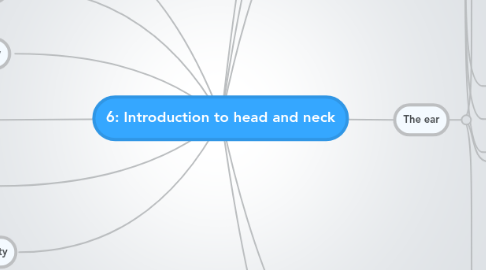
1. Pharynx
1.1. nasopharyx
1.1.1. has opening of auditory tube and adenoid
1.2. oropharynx
1.2.1. has the palatine tonsils
1.3. laryngopharynx
1.3.1. has the piriform fossa
2. Larynx
2.1. paired cartilages
2.1.1. arytenoid
2.1.2. corniculate
2.1.3. cuneiform
2.2. unpaired cartilages
2.2.1. thyroid
2.2.2. cridoid
2.2.3. epiglottis
2.3. cavity of the larynx
2.3.1. from inlet to lower part of cricoid
2.3.2. 3 parts
2.3.2.1. vestibule of the larynx
2.3.2.2. middle part of the larynx
2.3.2.2.1. from vestibular folds to the vocal folds
2.3.2.3. lower part of the larynx
3. scalp
3.1. hairy soft tissue covering the cranial vault
3.2. from supraorbital margin to external occipital protuberance & superior nuchal line posteriorly
3.3. on each side extends to superior temporal line
3.4. 5 layers (SCALP
3.4.1. Skin
3.4.2. Connective tissue (superficial facia)
3.4.3. Aponeurosis (galea aponeurotica)
3.4.4. Loose areolar CT
3.4.5. Pericranium
4. Face
4.1. boundaries
4.1.1. superiorly: the hair line
4.1.2. inferiorly: chin and base of mandible
4.1.3. on each side: auricle
4.2. remember
4.2.1. forehead is common to scalp & face
4.2.2. shape is determined by underlying bones
4.2.3. No deep facia in the face
5. Oral cavity
5.1. 2 parts
5.1.1. oral cavity proper
5.1.2. vestibule
5.2. contents
5.2.1. alveolar processes and teeth
5.2.2. anterior 2/3 of the tongue
5.2.3. orifice of parotid gland in buccul mucosa (opposite to upper second molar)
5.2.4. orifice of submandibular dict
5.2.5. orifice of sublingual glands in anterior floor of the mouth
6. The nasal cavity
6.1. nasal septum divides it into right and left
6.1.1. floor
6.1.1.1. palatine process of maxilla
6.1.1.2. horizontal plate of palatine bone
6.1.2. roof
6.1.2.1. body of sphenoid
6.1.2.2. cribriform of ethmoid
6.1.2.3. nasal bone
6.1.2.4. nasal cartilages
6.1.3. medial wall = nasal septum
6.1.3.1. osteocartilaginous partition
6.1.3.2. covered by mucus membrane
6.1.3.3. formed by:
6.1.3.3.1. vertical plate of ethmoid bine
6.1.3.3.2. vomer
6.1.3.3.3. nasal crest of palatine bone
6.1.3.3.4. septal cartilage
6.1.4. lateral wall
6.1.4.1. superior, middle, inferior nasal conchae
6.1.4.2. area below each conchae is a meatus
6.1.4.3. openings
6.1.4.3.1. maxillary sinus (middle meatus)
6.1.4.3.2. frontal sinus (middle meatus via infundibulum)
6.1.4.3.3. sphenoid sinus
6.1.4.3.4. ethmoidal sinuses
6.1.4.3.5. nasolacrimal duct
7. Orbits
7.1. Eyelids
7.2. lacrimal apparatus
7.3. The extraocular muscles
7.3.1. Innervation
7.3.1.1. oculomotor nerve
7.3.1.2. abducent nerve
7.3.1.3. trochlear nerve
7.3.2. two obliques
7.3.2.1. superior
7.3.2.1.1. origine: posterior wall of the orbital cavity
7.3.2.1.2. insertion: superior wall of the eyeball
7.3.2.2. inferior
7.3.2.2.1. origine: floor of the orbit
7.3.2.2.2. insertion: lateral wall of the eyeball
7.3.3. four recti
7.3.3.1. origine: tendinous ring on the posterior wall of the orbital cavity
7.3.3.2. rectus superior
7.3.3.3. rectus inferior
7.3.3.4. rectus medial
7.3.3.5. rectus lateral
7.3.3.6. insertion: superior, inferior, lateral, medial walls of the eyeball respectively
7.4. orbital facia
7.5. conjunctiva
7.6. orbital fat
8. Triangles of the neck
8.1. divided by sternocledomastoid
8.2. anterior triangle
8.3. posterior triangle
9. blood supply
9.1. common carotid artery
9.1.1. internal carotid
9.1.2. external carotid
9.1.2.1. superior thyroid
9.1.2.2. ascending pharyngeal
9.1.2.3. lingual
9.1.2.4. facial
9.1.2.5. occipital
9.1.2.6. posterior auricular
9.1.2.7. superficial temporal
9.1.2.8. maxillary
9.2. veins
9.2.1. anterior jugular
9.2.2. external jugular
9.2.2.1. posterior auricular
9.2.2.2. superficial temporal
9.2.2.3. maxillary
9.2.2.4. retromendibular
9.2.2.5. posterior external jugular
9.2.2.6. transverse cervical
9.2.3. internal jugular
9.2.3.1. lingual
9.2.3.2. facial
9.2.3.3. superior thyroid
9.2.3.4. pharyngeal
9.2.4. New node
10. Innervation
10.1. cranial nerves mainly
10.2. sympathetic trunk (vasomotor mainly)
10.3. cervical plexus (C1-C4)
11. The ear
11.1. external ear
11.1.1. auricle
11.1.1.1. helix
11.1.1.2. lobule
11.1.1.3. concha
11.1.1.4. tragus
11.1.2. external auditory meatus
11.1.2.1. outer third id cartilaginous
11.1.2.2. medial two thirds are boney
11.1.2.3. has hair, sebaceous, and ceruminous glands
11.1.3. innervated by auriculotemporal and vagus nerves
11.2. middle ear (tympanic cavity)
11.2.1. Roof
11.2.1.1. tegmen typnai (tegmental wall)
11.2.2. floor
11.2.2.1. jugular floor
11.2.3. lateral
11.2.3.1. membranous wall
11.2.4. medial
11.2.4.1. labyrinthine
11.2.5. anterior
11.2.5.1. carotid wall
11.2.6. auditory tube leads to nasopharynx
11.2.7. Auditory ossicles
11.2.8. muscles of the ear
11.3. inner ear
11.3.1. bony labyrinth
11.3.1.1. vestibule
11.3.1.2. semicircular canals
11.3.1.3. cochlea
11.3.2. membranous labyrinth
11.3.2.1. utricle
11.3.2.2. saccule
11.3.2.3. semicircular ducts
11.3.2.4. ducts of cochlea
12. Head and neck skeleton
12.1. skull (28)
12.1.1. cranial (8)
12.1.1.1. 1 frontal
12.1.1.2. 2 parietal
12.1.1.3. 1 occipital
12.1.1.4. 2 temporal
12.1.1.4.1. petrous portion
12.1.1.5. 1 sphenoid
12.1.1.6. 1 ethmoid
12.1.2. facial (14)
12.1.2.1. 2 zygomatic
12.1.2.2. 2 maxillae
12.1.2.3. 2 nasal
12.1.2.4. 2 lacrimal
12.1.2.5. 1 vomer
12.1.2.6. 2 palatine
12.1.2.7. 2 inferior conchae
12.1.2.8. 1 mandible
12.2. cervical vertebrae (7)
12.2.1. transverse foramen
12.2.2. atypical c1 (atlas), c2 (axis), and c7
12.2.2.1. Atlas C1
12.2.2.1.1. ring of bone
12.2.2.1.2. lacking a spinous process or body
12.2.2.1.3. has superior facets for occipital condyles
12.2.2.2. Axis C2
12.2.2.2.1. has "Dens" or "odontoid process"
12.2.2.2.2. has bifid spinous process
12.2.2.2.3. pivotal movement at atlanto-axial joint (No, rotation)
12.2.2.3. C7 (vertebra prominens)
12.2.2.3.1. has long (not bifid) spinous process
12.2.2.3.2. small transverse foramen for vein only
12.2.3. typical c3,c4,c5,c6
12.2.3.1. small rectangular bodies
12.2.3.2. large and triangular vertebral foramen
12.2.3.3. short transverse processes with foramen
12.2.3.4. short and bifid spinous process
12.2.3.5. superior and inferior articular facet
12.3. Hyoid bone
12.3.1. slender, curved bone
12.3.2. located inferior to the skull (between mandible and larynx)
12.3.3. dose not articulate with any other bone
12.3.4. suspended by stylohyiod ligaments
12.3.5. consists of
12.3.5.1. body
12.3.5.2. greater horns
12.3.5.3. lesser horns
12.3.6. it's the base for attachment of muscles and ligaments of the tongue and larynx
