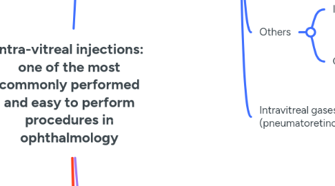
1. Drugs
1.1. Anti-VEGG
1.1.1. Pegaptinib (Macugen)
1.1.1.1. 1st FDA approved anti-VEGF for intra-vitreal injection in 2004
1.1.1.2. Approved for use for NAMD - not used now
1.1.1.3. RNA aptamer that binds and neutralizes VEGF165
1.1.2. Bevacizumab (Avastin)
1.1.2.1. off-label use for intra-vitreal injection
1.1.2.2. Recombinant humanized full length IgG1 antibody
1.1.2.3. non-specific VEGF inhibitor that binds to all VEGF-A isoforms and prevents their binding to endothelial cell receptors
1.1.2.4. Dose - 1.25mg/0.05 ml
1.1.2.5. Used commonly for - NAMD, PDR-DME, Maculae edema due to vascular occlusions
1.1.3. Ranibizumab (Lucentis, Accentrix, Razumab)
1.1.3.1. Recombinant humanized monoclonal antibody fragment
1.1.3.2. IgG1 light chin linked by a disulfide bond to a human igG1 Fab heavy chain, with murine anti-VEGF A complement determining regions
1.1.3.3. Produced in E coli
1.1.3.4. Molecular weight - 48kDa
1.1.3.5. Binds with high affinity to VEGF-A isoforms mainly - VEGF-165
1.1.3.6. Dose - 0.5 mg/ 0.05 ml
1.1.4. Aflibercept (Eyelea)
1.1.4.1. Recombinant fusion protein: chimeric protein that contains second Ig domain of human VEGFR2 both of which are fused to constant region of human IgG1
1.1.4.2. Produced in CHO cells
1.1.4.3. Molecular weight - 115 kDa
1.1.4.4. Binds to VEGF-A, B, C, D and Placental growth factor (PIGF)
1.1.4.5. Dose - 2mg/0.05 ml
1.1.5. Brolucizumab (Pagenax)
1.1.5.1. Humanized monoclonal single chain variable fragment antibody
1.1.5.2. Molecular weight of 26 kDa, binds and inhibits VEGF-A
1.1.5.3. FDA approved for NAMD
1.1.5.4. Dosing - 6 mg/0.05 ml
1.1.5.5. Dosing interval - 8-12 weeks - longer than other anti-VEGF agents
1.1.5.5.1. As it contains highest number of molecules per ANTI- VEGF ijection (11.2-12.3)
1.1.6. Faricimab
1.1.6.1. Bispecific antibody that targets VEGF-A and angiopoietin-2
1.1.6.2. FDA approved for NAMD and DME
1.1.6.3. Dosage - 6 mg/0.05 ml at 2 monthly intervals
1.2. Steroids
1.2.1. Injections
1.2.1.1. Triamcinilone
1.2.1.1.1. Dose - 2mg/ 0.05 ml
1.2.2. Implants
1.2.2.1. Dexamethasone
1.2.2.1.1. Ozurdex
1.2.2.2. Fluocinolone acetonide
1.2.2.2.1. Iluvien
1.2.2.2.2. Retisert
1.3. Anti-biotics
1.3.1. Used for endophthalmitis
1.3.1.1. Antibiotics
1.3.1.1.1. Individualized based on culture and sensitivity
1.3.1.2. Antifungals
1.3.1.2.1. Voriconazole - 0.1mg/0.1 ml
1.3.1.2.2. Amphotericin B - 5-10 micrograms/0.1 ml
1.4. Others
1.4.1. Immunosuppressive agents
1.4.1.1. Off label use to treat inflammation - uveitis: methotrexate, cyclosporine, tacrolimus
1.4.2. Chemotherapy
1.4.2.1. Used in retinoblastoma - Melphalan
1.5. Intravitreal gases (pneumatoretinopexy)
1.5.1. SF6
1.5.2. C3F8
2. Techniques
2.1. Equipment required
2.1.1. Sterile eye drape and eye towel, sterile eye speculum, sterile callipers, sterile cotton tipped applicator, 5% povidone iodine solutions/ eye drops, anaesthetic eye drops (proparacaine), sterile 1 ml syringe and sterile 29/30G needle, sterile gloves
2.2. Injection setting
2.2.1. clean room or OR
2.3. Sterility and other measures
2.3.1. 5% povidone iodine eye drops to be instilled 3 times at 5 minute intervals before the procedure and ocular surface to be cleaned with 5% povidone iodine solution, periocular skin to be cleaned with 10% povidone iodine solution
2.3.1.1. along with 5% povidone iodine eye drops topical anaesthetic eye drops should also be instilled
2.4. Distance of injection
2.4.1. In adults depends on lens status of patient - 3mm from limbus in aphakia. 3.5 mm from limbus in pseudophakia and 4 mm from limbus in phakic subjects
2.5. Injection procedure
2.5.1. after cleaning the ocular surface and periocular skin and applying the eye drape make a mark with the callipers on the sclera at the appropriate distance from the limbus
2.5.1.1. Injection/ needle to be directed towards the centre of the vitreous cavity/ perpendicular to the ocular surface
2.5.1.1.1. preferable to stabilize the needle hub with one hand and inject with the other hand - needle can be inserted completely till hub of the needle
2.6. Other tips
2.6.1. Avoid talking during the procedure - oral flora have been shown to be a source of endophthalmitis
2.6.2. Place the eye drape gently and avoid excess manipulation of the lids to prevent expression of meibomian gland secretions
2.6.3. Immediately after the procedure on table check for:
2.6.3.1. Perception of light
2.6.3.2. Intra-ocular pressure with a cotton tipped applicator - if high perform AC paracentesis
2.6.4. Avoid injecting both eyes on the same day where possible
2.6.5. Tell the patient to avoid eye rubbing, avoid a head bath for 48 hours and avoid swimming for 3 days
2.7. ROP injections
2.7.1. half dose injection or 1/3rd dose injection
2.7.2. injected at 1 mm from limbus
2.7.2.1. both eyes injected in same setting
2.7.2.1.1. Treat each eye as a new case with separate scrubbing and draping
2.7.3. needle inserted completely till hub and directed perpendicular to table/ floor and not perpendicular to ocular surface
2.7.4. eye massage with cotton tipped applicators to be done immediately after injection to reduce IOP
3. ComplicationsComplications
3.1. retinal tear
3.2. Endophthalmitis- low incidence
3.2.1. 0.04-1.4%
3.3. Secondary Glaucoma
3.4. Anti VEGF crunch syndrome
3.4.1. Most common with bevacizumab
3.4.1.1. usually seen 2-6 weeks post AntiVEGF
3.5. Vitreous haemorrhage
3.6. Accidental injury to lens capsule
4. Miscellaneous and updates
4.1. MARINA study- Ranibizumab beneficial in minimally classic/Occult CNVM
4.2. ANCHOR trial- Ranibizumab superior to PDT in subfoveal classic CNVM
4.3. RESTORE- Ranibizumab ,Ranibizumab +laser bettter than Laser lone
4.4. DRCR, net
4.4.1. Protocol B- Laser photocoagulation>IVTA
4.4.2. Protocol I- IV Ranibizumab + Prompt lase better than prompt laser alone in center involving DME
4.4.3. Protocol S- Ranibizumab is as effective as PRP in PDR
4.4.4. Protocol T-aflibercept superior than ranibizumab and bevacizumab in DME
