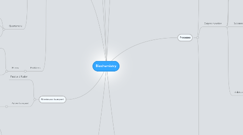
1. Protein structure
1.1. Levels
1.1.1. Primary
1.1.1.1. Aminoacid sequence
1.1.1.1.1. Aminoacids
1.1.2. Secondary
1.1.2.1. Alpha helix
1.1.2.1.1. Right hand coiled
1.1.2.1.2. Backbone NH H links to C=O
1.1.2.1.3. 3.6 resudues per turn
1.1.2.2. Beta sheet
1.1.2.2.1. Lateral connection
1.1.2.2.2. At least 2-3 bonds per line
1.1.2.2.3. Configurations
1.1.2.3. No covalent bonds
1.1.3. Tertiary
1.1.3.1. 3D structure
1.1.3.2. Non-specific hydrophoibic interactions
1.1.3.2.1. Salt bridges
1.1.3.2.2. H bonds
1.1.3.2.3. Disulfide bonds
1.1.3.2.4. Tight packing of side chains
1.1.4. Quarternaru
1.1.4.1. Multi tertiary units
1.1.4.2. Same noncovalent bonds as tertiary
1.1.4.3. Multiple polypeptides
1.1.4.3.1. Multimere
1.1.4.3.2. Di/trimer etc
1.2. Problems
1.2.1. Prions
1.2.1.1. Transmissable confirmational changes
1.2.1.2. PrP
1.2.1.2.1. PrP(c) is ok
1.2.1.2.2. PrP(sc) is not ok
2. Specific proteins
2.1. Hemoglobin
2.1.1. Hemoglobin saturation curve
2.1.1.1. CO2 decreases O2 affinity
2.1.1.2. BPG lowers O2 affinity
2.1.1.3. First O2 molecule binds
2.1.1.3.1. heme Fe ion is drawn into the porphyrin ring plane
2.1.1.4. H+ promotes dissociation
2.1.1.5. Deoxyhemoglobin
2.1.1.5.1. Higher H+ affinity
2.1.1.5.2. Some CO2 transported directly
2.1.1.6. Fetal blood has lower BPG affinity
2.1.1.6.1. Hogher O2 affinity
2.1.2. Structure
2.1.2.1. 2 a-units
2.1.2.2. 2 b-units
2.1.2.3. Each unit has heme group with Fe
2.1.3. Trouble
2.1.3.1. Sicle cell disease
2.1.3.1.1. Advangates
2.1.3.1.2. Characteristics
2.2. Myoglobin
2.2.1. Behavior
2.2.1.1. Binds 1 O2
2.2.1.2. Traditional Michaelis-menten graph
2.2.1.3. Oxygen storage in muscle
2.3. Collagen
2.3.1. Structure
2.3.1.1. Connective tissue
2.3.2. Prolyl hydroxylase
2.3.2.1. Needs vit C to function
2.3.2.1.1. Cofactor to keep Fe++ in reduced state
2.3.2.2. Involved in collagen synthesis
3. Genetics
3.1. Gene expression
3.1.1. Transcription
3.1.1.1. Phases
3.1.1.1.1. Pre-initiation
3.1.1.1.2. Initiation
3.1.1.1.3. Promoter clearence
3.1.1.1.4. Elongation
3.1.1.1.5. Termination
3.1.1.2. Steps
3.1.1.2.1. DNA to mRNA
3.1.1.2.2. Recognition of promoter (TATAAT)
3.1.1.2.3. RNA-polymerase binds
3.1.1.2.4. Helicase enzymes make transcription bubble
3.1.1.2.5. RNA polymerase runs
3.1.1.2.6. 3'UTR poly-A tail and a 5'UTR cap
3.1.2. Translation
3.1.2.1. Initiation
3.1.2.1.1. 30S ribosome complex binds
3.1.2.1.2. IF-2 delivers tRNA-fMet into P site
3.1.2.1.3. 50S subunit binds
3.1.2.2. Elongation
3.1.2.2.1. Complex shit, will add if bored
3.1.2.3. Termination
3.1.2.3.1. Stop codon found
3.1.2.3.2. RF-/RF2 +RF-3:GTP binds to A site
3.1.2.3.3. Confirmational jumbo
3.1.2.3.4. Dissociation
3.2. RNA
3.2.1. Can cleave own backbone
3.2.1.1. 2' hydroxyl oxygen attacks the phosphodiester bond
3.2.2. Uracil
3.2.2.1. Replaces thymine in RNA
3.2.2.2. Uracil methylation results in Thymine
3.2.2.3. Deaminated cytosine is uracil
3.2.2.3.1. Uracil DNA glycosilate fixes
3.2.3. Evolved before DNA
3.2.4. Types
3.2.4.1. Mainstream
3.2.4.1.1. mRNA
3.2.4.1.2. tRNA
3.2.4.1.3. rRNA
3.2.4.1.4. ncRNA
3.2.4.2. Lesser known
3.2.4.2.1. miRNA
3.2.4.2.2. siRNA
3.2.4.2.3. piRNA
3.2.4.2.4. snRNA
3.2.4.2.5. snoRNA
3.2.4.2.6. dsRNA
3.3. DNA
3.3.1. PCR
3.3.1.1. Step 1
3.3.1.1.1. Isolate target sequence
3.3.1.1.2. Heat to 95 celcius
3.3.1.1.3. Cool to 70
3.3.1.2. Step 2
3.3.1.2.1. Add 1000 fold excess primer
3.3.1.3. Step 3
3.3.1.3.1. (Taq) DNA polymerase
3.3.1.3.2. dTTP, dATP, dGTP, dCTP
3.3.1.4. Step 4
3.3.1.4.1. Heat to 95
3.3.1.4.2. Cool to 70
3.3.1.5. Step 5
3.3.1.5.1. Repeat 4
3.4. Forensic PCR
3.4.1. DNA profiling
3.4.1.1. VNTRs
3.4.1.1.1. variable number tandem repeats
3.4.1.2. STR
3.4.1.2.1. particularly short tandem repeats
4. Phospholipids
4.1. Structure
4.1.1. 1 fatty acid tails
4.1.1.1. COO attached to glycerol
4.1.1.2. On 1 and 2
4.1.2. Glycerol
4.1.3. Phosphate group
4.1.3.1. G-OP(=O)(O-)-O-topgroup
4.1.3.2. On 3
4.1.4. Top group of sorts
4.1.4.1. Often choline
5. Membrane transport
5.1. Passive diffusion
5.2. Active transport
5.2.1. Transporter proteins
5.2.1.1. Primary
5.2.1.1.1. Pump
5.2.1.1.2. ATP
5.2.1.2. Secondary
5.2.1.2.1. Uniport
5.2.1.2.2. Cotransport
5.2.1.2.3. Gradient
6. pH
6.1. Hyperventilation
6.1.1. Increased gass/lung contact
6.1.2. Lower CO2
6.1.2.1. Lower H2CO3
6.1.2.1.1. Lower H+ + HCO3-
6.1.2.2. Hb affinity for O2 increases
6.1.2.2.1. Lower O2
6.2. Formulas
6.2.1. pH = -Log[H+]
6.2.2. [H+] = 10^-pH
6.2.3. [H+] = ([HA]Ka)/[A-]
6.2.4. [H+][OH-]=10^-14 M
6.2.5. Weak acid
6.2.5.1. Ka = [H+][A-] / [HA]
6.2.5.1.1. Ka low = weak acid
6.2.5.1.2. pKa = -log(Ka)
7. Processes
7.1. Blood clotting
7.1.1. Vitamin K
7.1.1.1. Function
7.1.1.1.1. Add carboxylic acid group to a glutamate amino acid residue
7.1.1.1.2. Usage
7.1.1.1.3. Reduction and subsequent reoxidation of vitamin K coupled with carboxylation of Glu is called the vitamin K cycle
7.2. Enzyme function
7.2.1. Features
7.2.1.1. Protein/RNA catalysts
7.2.1.2. Specificity
7.2.1.3. Regulation
7.2.1.3.1. Enzyme expression
7.2.1.3.2. Enhancers
7.2.1.3.3. Inhibitors
7.2.1.4. Catalytic power
7.2.1.4.1. Lower activation energy
7.2.2. Additional components
7.2.2.1. Cofactors
7.2.2.2. Coenzymes
7.2.2.2.1. If tightly bound
7.2.3. Calculations
7.2.3.1. v = d[A]/dt = K[A]
7.2.3.2. A+B > P+Q
7.2.3.2.1. v= dA/dt = dB / dt = dP /dt = dQ / dt
7.2.3.3. ES <k1 / k-1 > E+S
7.2.3.3.1. (k-1)[ES]<>(k1)[E][S]
7.2.3.3.2. Ks=[ES]/[E][S] = (k-1)/(k1)
7.2.3.4. E+S <k1/k-1> ES >k2> E+P
7.2.3.4.1. Km = (K1+K2)/(K-1)
7.2.3.4.2. v =
7.2.4. Substrate binding
7.2.4.1. H bond
7.2.4.2. Ionic
7.2.4.3. Vd Waals
7.2.5. Inhibitors
7.2.5.1. Effects
7.2.5.1.1. Km
7.2.5.1.2. Vmax
7.2.5.2. Reversible
7.2.5.2.1. Competitive
7.2.5.2.2. Noncompetitive
7.2.5.2.3. Uncompetitive
7.2.5.3. Irreversible
7.2.5.3.1. Suicide substrate
7.3. Glycogen
7.3.1. Glycogenolysis
7.3.1.1. Glycogen(n residues) + Pi <> glycogen(n-1 residues) + glucose-1-phosphate
7.3.1.1.1. 1. Glycogen phosphatase phosphorilates a-1-4
7.3.1.1.2. 2. G1P to G6P
7.3.2. Glycogenesis
7.3.2.1. 1. Glucose to G6P
7.3.2.1.1. Glucokinase
7.3.2.1.2. Hexokinase
7.3.2.2. 2. G6P to G1P
7.3.2.2.1. Phosphoglucomutase
7.3.2.3. 3. G1P to UDP-Glucose
7.3.2.3.1. Uridyl Transferase
7.3.2.3.2. Pyrophosphate formed
7.3.2.4. 4. Chain assembly
7.3.2.4.1. Glycogen synthase
7.3.2.5. 5. Branching
7.3.2.5.1. amylo-α(1:4)→α(1:6)transglycosylase
8. Polysaccharides
8.1. Linked glucose units
8.1.1. Starch
8.1.1.1. a-1-4 Glycocidic bonds
8.1.2. Cellulose
8.1.2.1. b-1-4 Glycocidic bonds
8.1.3. Glycogen
8.1.3.1. a-1-4 linkages
8.1.3.2. a-1-6 linksge branches
8.1.4. Dextrans
8.1.4.1. a-1-6 linkages
8.1.4.2. a1-3 branches
8.2. Linked Agarobiose units
8.2.1. Agarose
8.2.1.1. 1-4 bonds
8.2.2. = D-galactose + 3,6-anhydro-L-galactopyranose
8.3. Linked N-Acetylglucosamine
8.3.1. Chitin
8.3.1.1. b-1-4 linkage
8.3.1.2. Structure similar to cellulose
