

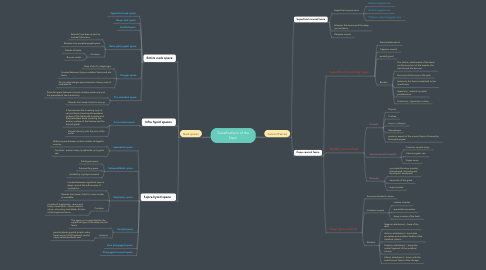
1. Cervical Fascias
1.1. Superficial cervical fascia
1.1.1. Platysma muscle
1.1.2. Superficial cervical veins
1.1.2.1. External jugular vein
1.1.2.2. Anterior jugular vein
1.1.2.3. Posterior external jagular vein
1.1.3. between the dermis and the deep cervical fascia
1.2. Deep cervical fascia
1.2.1. Superficial (investing) layer
1.2.1.1. Sternocleidomatoid
1.2.1.2. Trapezius muscle
1.2.1.3. parotid gland
1.2.1.4. Borders
1.2.1.4.1. The inferior attachments of the fascia are the acromion of the scapula, the clavicle and the sternum.
1.2.1.4.2. Surround all structure in the neck
1.2.1.4.3. Anteriorly this fascia is attached to the hyoid bone.
1.2.1.4.4. Superiorly , external occipital protuberance
1.2.1.4.5. Posteriorly , ligmentum nuchae
1.2.2. Middle (pretracheal)
1.2.2.1. Visceral
1.2.2.1.1. Thyroid
1.2.2.1.2. Trachea
1.2.2.1.3. Larynx + pharynx
1.2.2.1.4. Oesophagus
1.2.2.1.5. posterior aspect of the visceral fascia is formed by Buccopharyngeal
1.2.2.2. Vascular(carotid sheath)
1.2.2.2.1. Common carotid artery
1.2.2.2.2. Internal jugular vein
1.2.2.2.3. Vagus nerve
1.2.2.3. Muscular
1.2.2.3.1. surrounds the strap muscles (sternohyoid, sternothyroid, thyrohyoid, omohyoid)
1.2.2.3.2. adventitia of the great
1.2.2.3.3. strap muscles
1.2.3. Deep (preverebral)
1.2.3.1. Surround vertebral column
1.2.3.2. Vertebral muscle
1.2.3.2.1. scalene muscles
1.2.3.2.2. prevertebral muscles
1.2.3.2.3. deep muscles of the back.
1.2.3.3. Borders
1.2.3.3.1. Superior attachment – base of the skull.
1.2.3.3.2. Anterior attachment – transverse processes and vertebral bodies of the vertebral column.
1.2.3.3.3. Posterior attachment – along the nuchal ligament of the vertebral column
1.2.3.3.4. Inferior attachment – fusion with the endothoracic fascia of the ribcage.
2. Neck spaces
2.1. Entire neck space
2.1.1. Superficial neck space
2.1.2. Deep neck space
2.1.3. Carotid space
2.1.4. Retro pharyngeal space
2.1.4.1. Extends from base of skull to tracheal bifurcation
2.1.4.2. Between two parapharyngeal space
2.1.4.3. Contains
2.1.4.3.1. Glands of Henle
2.1.4.3.2. Rouvier nodes
2.1.5. Danger space
2.1.5.1. Base of skull to diaphragm
2.1.5.2. Located between the pre vertebral fascia and alar fascia
2.1.5.3. This is called danger space because of easy route of mediastinitis
2.1.6. Pre vertebral space
2.1.6.1. Potential space between cervical vertebra posteriorly and the prevertebral fascia anteriorly
2.1.6.2. Extends from base of skull to coccyx
2.2. Infra hyoid spaces
2.2.1. Pre tracheal space
2.2.1.1. It lies between the Investing layer of cervical fascia (covering the posterior surface of the Infrahyold muscles) and the pretracheal fascia (covering the anterior surface of the trachea and the thyroid gland).
2.2.1.2. blends Inferiorly with the arch of the aortal.
2.3. Supra hyoid space
2.3.1. Submental space
2.3.1.1. Midline space between anterior bellies of digastric muscles
2.3.1.2. Contents - areolar tissue, lymphnode, ant jugular vein
2.3.2. Submandibular space
2.3.2.1. Sublingual space
2.3.2.2. Submaxillary space
2.3.2.3. divided by mylohyoid muscle
2.3.3. Masticator space
2.3.3.1. Located between superficial layer of deep cervical fascia & muscles of mastication
2.3.3.2. Extends from base of skull to lower border of mandible
2.3.3.3. Contains
2.3.3.3.1. muscles of mastication, ramus and body of mandible , inferior alveolar nerve, vein,artery mandibular division of the trigeminal nerve
2.3.4. Parotid space
2.3.4.1. The space is circumscribed by the superficial layer of the deep cervical fascia
2.3.4.2. contents
2.3.4.2.1. parotid glands parotid lymph nodes facial nerve (CN VII) external carotid artery retromandibular vein
2.3.5. Para pharyngeal space
2.3.6. Pharyngeal mucosal space