

1. Introduction on L.A
1.1. Definition of L.A
1.1.1. (a loss of sensation in a circumscribed area of the body), (caused by a depression of excitation in nerve endings), or (inhibition of, the conduction process in peripheral nerves)
1.2. methods of inducing Local Anesthesia
1.2.1. Mechanical Trauma
1.2.2. Low Temperature
1.2.3. Anoxia
1.2.4. Chemical Irritants
1.2.5. Neurolytic agents such as alcohol & phenol
1.2.6. Chemical agents such as local anesthetics
1.3. Desirable properties of Local Anesthetics
1.3.1. should not be irritating to the tissue
1.3.2. not cause permanent nerve structure damage
1.3.3. Its systemic toxicity should be low
1.3.4. must be effective as an Injection or if applied topically
1.3.5. Short time of onset
1.3.6. Long duration of action , but not too long
1.3.7. have sufficient potency without use of concentrated solutions
1.3.8. not have allergic reaction potential
1.3.9. should be stable in solution & undergo readily biotransformation in the body
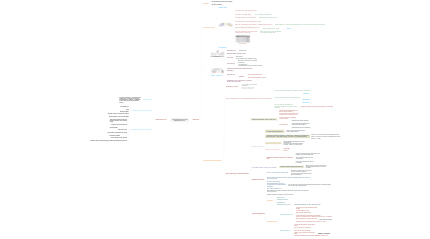
2. The Neuron
2.1. Definition
2.1.1. the structural unit of the nervous system
2.1.2. It transmits messages between the CNS & all parts of the body
2.2. portion of the nerve
2.2.1. Dendritic zone
2.2.2. The axon
2.2.2.1. It’s a nerve fibre, long cylindrical neural cytoplasm
2.2.2.2. encased in thin sheath called?
2.2.2.2.1. nerve membrane or Axolemma.
2.2.2.3. changes within the nerve membrane is what is responsible for ?
2.2.2.3.1. Responsible for the Sensory nerve excitability & Conduction
2.2.2.4. nerve membrane is 70 to 80 Angstrom unit thick
2.2.2.5. Axon can be covered by Myelin sheath ( an insulating Lipid rich layer )
2.2.2.5.1. Myelin sheaths are a actually a specialized form of (Schwan cells) which wrap the nerve fibres
2.2.2.6. there are Myelinated & Non Myelinated nerves
2.2.2.6.1. Myelinated nerves conduct faster than Non-myelinated nerves , why?
2.2.2.7. There are constrictions located at regular intervals which is called as???
2.2.2.7.1. Node of Ranvier as it is the gap between two adjoining Schwann cells
2.2.2.8. Classifications
2.2.3. The cell body
2.3. Types
2.3.1. Sensory ( afferent )
2.3.1.1. Dendritic zone
2.3.1.1.1. responds to the stimulus & produce an impulse which is transmitted along the axon
2.3.1.2. Pseudounipolar & shorter axon
2.3.1.3. The axon
2.3.1.3.1. thin cable like
2.3.1.3.2. can be sometimes upto 100 –200 cm long
2.3.1.4. The cell body
2.3.1.4.1. NOT involved in the process of impulse transmission
2.3.1.4.2. it’s function is to: provide metabolic support for the sensory neuron
2.3.2. Motor ( efferent )
2.3.2.1. conduct impulses from the CNS toward the periphery
2.3.2.2. The cell body
2.3.2.2.1. between the Axon & the Dendrites
2.3.2.2.2. Functions
2.3.2.3. multipolar & has a longer axon compared to the sensory neuron
2.4. Physiology of Peripheral Nerves
2.4.1. The function of nerve
2.4.1.1. is to carry messages from one part of the body to another.
2.4.1.2. The messages are in the form of Electrical Action potential also called as Impulses
2.4.2. Action potentials are due to transient (Depolarization)that occur in the nerve membrane
2.4.2.1. ((due to increase of permeability of Sodium ions into the membrane))
2.4.2.2. Impulses are generated by the following Stimuli:
2.4.2.2.1. Chemical
2.4.2.2.2. Thermal
2.4.2.2.3. Mechanical
2.4.2.2.4. Electrical
2.4.2.3. Once an Impulse is generated the Amplitude, & shape of the Impulse remains constant
2.4.2.3.1. regardless of changes in the quality of the stimulus or its strength
2.4.3. Electro-Physiology of Nerve Conduction
2.4.3.1. 1-Resting Potential: -70mV ( milli-Volt )
2.4.3.1.1. negative electrical potential that exists across the nerve membrane.
2.4.3.1.2. due to the differing concentration of ions on either side of membrane (In & out)
2.4.3.1.3. The interior of the nerve is Negative relative to exterior
2.4.3.1.4. nerve membrane is:
2.4.3.2. Stimulants stimulate :
2.4.3.2.1. 2-initial slow Depolarization
2.4.3.2.2. 3-electrical potential reaches a critical level= extremely rapid phase of Depolarization , this called (Firing Threshold or Threshold Potential)
2.4.3.2.3. 4-Repolarization occurs
2.4.3.2.4. Phase 2,3 and 4 require 1 ms
2.4.3.2.5. Sequence of events of phase 2,3,4 depends on??
2.4.3.3. for a brief period the nerve is unable to respond to another stimulus this is called
2.4.3.3.1. 5-Refractory Period(hyperpolarisation)
2.4.3.4. Membrane Channels
2.4.3.4.1. There are specific channels for the ions like Na & K
2.4.3.4.2. Sodium channels also have the ability to change the shape depending on the charges of the nerve membrane
2.4.3.4.3. membrane permeability is due to the presence of these channels
2.4.3.4.4. During Depolarization Na ions pass through due to the changes within the membrane
2.4.3.4.5. Na + water is difficult to pass
2.4.3.5. Impulse Propagation
2.4.3.5.1. After initiation of the action potential by a stimulus the impulse must move along the surface of the axon
2.4.3.5.2. Impulse propagation can happen in only one direction
2.4.3.5.3. Sequence:
2.4.3.5.4. Impulse Spread