
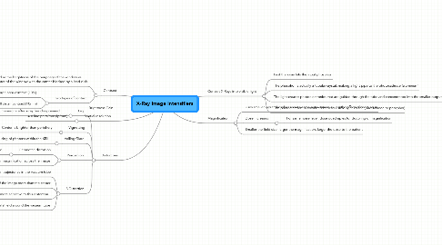
1. Contrast
1.1. Defined as the brightness of the periphery of the window vs the center of the window with the center blocked by a lead disk.
1.2. Two types of contast
1.2.1. large area contrast (10%)
1.2.1.1. 10:1 to 30:1 is typical
1.2.2. small detail contrast (10mm)
1.2.2.1. 15:1 to 35:1 is typical
2. Brightness Gain
3. Spatial resolution
3.1. 4-6 line pairs/mm (lp/mm)
4. Distortions
4.1. Lag
4.1.1. Persistance of lumenescne after x-ray has disappeared
4.1.1.1. Not as big a deal these days - older II's had 30-40ms lag, new ones have 1ms.
4.1.1.2. "lag" in modern fluoroscopic systems is more likely caused by the CCTV system than the XRII
4.2. Vignetting
4.2.1. Center is brighter than periphery
4.2.1.1. --> Center has higher resolution, less distortion, increased brightness
4.3. Veiling Glare
4.3.1. Scattering of light, defocusing of photons within the XRII
4.3.1.1. X-ray glare, phosphor spreading, electron scatter all contribute
4.4. Pincushion
4.4.1. Geometric distortion
4.4.1.1. New node
4.4.2. Nonlinear magnification across the image
4.5. S Distortion
4.5.1. Caused by external magnetic fields affecting photoelectron trajectories in the vacuum tube
4.5.2. Affects the perimeter of the image more than the center
4.5.3. Larger XRII's are more sensitive to this distortion
4.5.4. Manufacturers include a highly conducting metal shield around the vacuum tube
5. Convert X-Rays into visible light
5.1. First the x-ray hits the input phosphor
5.2. The phosphor is actually a tubular crystal, making a light pipe to the photocathode (aluminum)
5.3. The light creates photoelectronics that are guided through the tube and concentrated into the smaller output opening via EM fields created by electrodes along the walls of the tube
5.3.1. It is in this stage that the distortions occur through influence of external magnetic fields on the guidance fields
5.4. The photoelectron is converted back to visible light by the (photoanode or phosphor)
6. Magnification
6.1. Uses smaller input phosphor area to same output area, giving the effect of zooming in
6.2. Dose increases
6.2.1. For same noise level, dose quadruples for doubling of magnification