

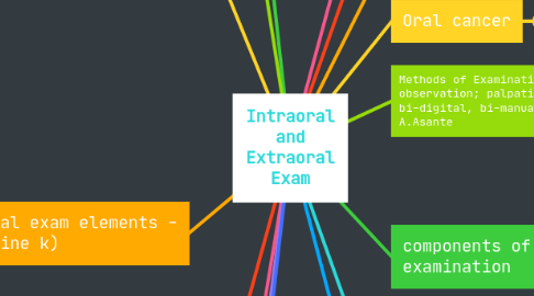
Intraoral and Extraoral Exam
by MikeWendy Herndon
1. Physical Characteristics
1.1. Size & shape
2. Intraoral exam
2.1. Soft tissue, teeth, and gums, hard palate, soft palate, alveolar and vestibular mucosa, floor of mouth, TMJ R.Harrison
2.2. Palpatation by feeling for tenderness or swelling in the mouth, and lymph nodes under the jaw or neck area R.Harrison
2.3. Mouth mirror is used for invisibility and inspection, and explorer and probe are used for detecting cavities or rough surfaces R.Harrison
3. Kinds of lesions
3.1. elevated
3.1.1. Blisterform (vesicle, pustule, bulla) - Gabby T
3.1.2. Nonlblisterform (papule, nodule, tumor, plaque) - Gabby T
3.1.3. Depressed lesions (ulcer, erosion) - Gabby T
3.1.4. Flat lesions (macule) - Gabby T
4. extraoral exam elements - (katherine k)
4.1. overall appearance
4.2. face
4.2.1. visual
4.3. skin
4.3.1. dry/lesions
4.4. nodes
4.4.1. palpation
4.5. TMJ
4.5.1. feeling the joint from palpation
4.6. lips
5. Palpation: the evaluation of a lesion by feeling it with fingers to determine the texture of the area. (Natarsha B)
5.1. Soft
5.2. Firm
5.3. Semifirm
5.4. Fluid filled
6. Documentation (Grace D)
6.1. Very necessary to accurately and precisely document every aspect of the extra and intra oral exam (Grace D)
6.2. Use dental terms or normal descriptive words to describe what you see (Grace D)
6.2.1. Examples: Corrugated, lobulated, pallor, vascular, fissured, linea alba, etc. (Grace D)
7. Tb
8. Methods for Examination (Lisely G.)
8.1. Palpation- using the sense of touch through tissue manipulation or pressure on an area with gloved finger of one hand or both
8.1.1. Bimanual palpayion
8.1.2. Bilateral palpation
9. palpation intraoral imaging
9.1. Anatomy teeth gongiva tongue palate salaivary glands cheeks floor of the mouth
10. Examinations
10.1. Visual insspection
11. Necessary as part of a comprehensive exam
12. Used to detect abnormalities
12.1. palpation
12.1.1. Observation
12.1.1.1. Olfaction