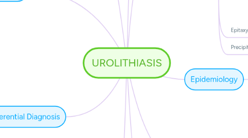
1. Differential Diagnosis
1.1. Appendicitis
1.2. Papillary necrosis
1.3. Pyelonephritis
1.4. Tumor
2. Complication
2.1. Hydronephrosis
2.2. Sepsis
2.3. Kidney Damage
2.4. Urinary Retention
2.5. Urinary Tract Obstruction
3. Treatment
3.1. Pharmacology
3.1.1. Tamsulosin
3.1.2. Alkali Treatment
3.1.3. Opioid
3.1.4. NSAIDS
3.1.5. Allupurinol
3.1.6. Thiazide Diuretics
3.2. Non Pharmacology
3.2.1. Nutritional Aspect
3.2.2. Open Surgery
3.2.3. Laproscopic Surgery
3.2.4. Ureterorenoscopy
3.2.5. Percutaneous Nephrolithotomy
3.2.6. Exrracorporeal Shockwave Lithrioscopy
4. Diagnosis and Test
4.1. Imaging Test
4.1.1. Retrograde Pyelography
4.1.2. CT SCAN
4.1.3. IV pyelography
4.1.4. Plain Film Radiography
4.1.5. Abdominal USG
4.2. Blood Test
4.2.1. BUN
4.2.2. GFR
4.2.3. Serum Ceratinine
4.3. Urinalysis
4.3.1. WBC
4.3.2. Urinary Crystal
4.3.3. Hematuria
4.4. History Taking
5. Prognosis
5.1. <5 mm (80% pass)
5.2. >1 cm need intervention
5.3. >5 mm (<50% pass)
6. RIsk factors
6.1. External
6.1.1. Climate and Geography
6.1.2. Diet and Food Intake
6.1.3. Occupation
6.2. Internal
6.2.1. Race & Ethnicity
6.2.2. Age & Gender
6.2.3. Genetics
7. Epidemiology
7.1. Climate and Socio economic
7.2. 0,1-0,4% of the population
7.3. 1-5% in Asia
7.4. 2-13% in develop country
8. Pathogenesis
8.1. Types of Stone
8.1.1. Calcium Phosphate
8.1.2. Indinavir
8.1.3. Uric Acid
8.1.4. Struvite
8.1.5. Cyesteine
8.1.6. Calcium Oxalate
8.2. Renal Colic (worst pain radiates to groin)
8.2.1. Contraction ureteral smooth muscle
8.2.2. Stimuli of Nociceptor
8.2.3. PG E-2
8.2.4. C Fibers

