

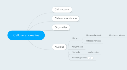
1. Cell patterns
1.1. Cell shape
1.1.1. Cuboidal cells
1.1.2. Columnar cells
1.1.2.1. Clear pale cytoplasm
1.1.3. Polygonal
1.1.4. Round cells
1.1.4.1. Small round cells
1.1.4.1.1. Small myoid cells or plasmacytoid cells
1.1.4.1.2. Small round blue cells
1.1.4.2. Large round cells
1.1.4.2.1. Large myoid cells
1.1.5. Small cells
1.1.5.1. Eccentric, crescentic nuclei
1.1.6. Large cells
1.1.6.1. Ballon cells
1.1.6.2. Abundant pale cytoplasm
1.1.6.3. Abundant eosinophilic cytoplasm
1.1.7. Stellate cells
1.1.8. Spindle cells / Fusiform cells
1.1.8.1. Fibroblastic/Myofibroblastic cells
1.1.8.1.1. Fibrohistiocytic cells
1.1.8.1.2. Fibroblastic cells
1.1.8.1.3. Myofibroblastoma
1.1.8.1.4. Atypical fibroxanthoma
1.1.8.1.5. Myofibroblastic sarcoma
1.1.8.1.6. Fibrous tumor of soft tissue
1.1.8.2. Schwann cells
1.1.8.2.1. Schwannoma
1.1.8.2.2. Neurilemmoma
1.1.8.3. Meningioma
1.1.8.4. Adipocytic cells
1.1.8.5. Smooth muscle cells
1.1.8.5.1. Leiomyoma
1.1.8.5.2. Leiomyosarcoma
1.1.8.6. Striated muscle cells
1.1.8.6.1. Rhabdomyosarcoma
1.1.8.7. Melanocytic cells
1.1.8.7.1. Spitz naevus
1.1.8.7.2. Melanoma
1.1.8.7.3. Melanocytoma
1.1.8.8. Epithelial cells
1.1.8.8.1. Sarcomatoid carcinoma / Carcinosarcoma
1.1.8.9. Endothelial cells
1.1.8.9.1. Kaposi sarcoma
1.1.8.9.2. Angiosarcoma
1.1.8.9.3. Kaposiform hemangioendothlioma
1.1.8.10. Cajal cells
1.1.8.10.1. GIST
1.1.8.11. Interdigitating dendritic cell sarcoma
1.1.9. Giant multinucleate cells
1.1.10. Cells with giant nuclei
1.2. Cell type
1.2.1. Columnar
1.2.2. Lipocytic
1.2.3. Malpighian
1.2.4. Rhabdoid
1.2.5. Fibroblastic/myofibroblastic
1.2.6. Sebaceous or hibernomatous
1.2.7. Oncocytic / Oxyphilic
1.2.8. Apocrine cells
1.3. Cell appearance
1.3.1. Cytoplasm
1.3.1.1. Scanty
1.3.1.1.1. Small round blue cells
1.3.1.2. Abundant
1.3.1.2.1. Eosinophilic
1.3.1.2.2. Clear / Pale
1.3.1.3. Abundant cytoplasm
1.3.1.3.1. Eosinophilic / granular
1.3.1.3.2. Clear cytoplasm
1.3.1.3.3. In mucinous material
1.3.1.4. Bodies
1.3.1.4.1. Spherical
1.3.1.4.2. Eosinophilic
1.3.1.4.3. Hyaline
1.3.1.4.4. Rhabdoid
1.3.2. Nuclei
1.3.2.1. Multinucleate cells
1.3.2.1.1. Frequent
1.3.2.1.2. Rare
1.3.2.2. Size
1.3.2.2.1. Small
1.3.2.2.2. Medium
1.3.2.2.3. Large
1.3.2.2.4. Monstruous
1.3.2.3. Shape
1.3.2.3.1. Round
1.3.2.3.2. Angulous
1.3.2.3.3. Irregular
1.3.2.3.4. Pleomorphic
1.4. Cell appearance
1.4.1. Cell size
1.4.1.1. Small size
1.4.1.1.1. Small cells
1.4.1.2. Medium size
1.4.1.3. Large size
1.4.1.4. Giant cell
1.4.1.4.1. Medullary thyroid tumor
1.4.2. Cell shape
1.4.2.1. Round cell
1.4.2.2. Polygonal cell
1.4.2.2.1. Polygonal or round cells
1.4.2.3. Fusiform cell
1.4.2.3.1. Spindle cells, irregularly arranged
1.4.3. Sebaceous cells - Hibernomatous cells
1.4.3.1. Sertoli cell tumor
1.5. Nucleus
1.5.1. Chromatin
1.5.1.1. Coarse
1.5.1.2. Fine
1.5.2. Nucleole
1.5.2.1. Marked
1.5.3. Shape
1.5.3.1. Crescentic
1.5.4. Position
1.5.4.1. Central
1.5.4.2. Eccentric
1.5.5. Size
1.5.5.1. Small
1.5.5.2. Medium
1.5.5.3. Large
1.5.5.4. Giant
2. Cellular membrane
3. Organelles
4. Nucleus
4.1. Mitosis
4.1.1. Abnormal mitosis
4.1.1.1. Multipolar mitosis
4.1.2. Mitoses increase
4.2. Karyorrhexis
4.3. Nucleole
4.3.1. Nucleolation