
1. Magnetic Resonance Imaging or MRI
1.1. How it works
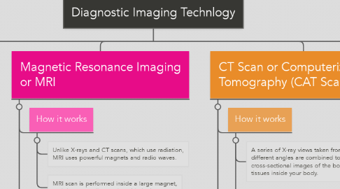
1.1.1. Unlike X-rays and CT scans, which use radiation, MRI uses powerful magnets and radio waves.
1.1.2. MRI scan is performed inside a large magnet, and the person lies on the table in the center.
1.1.2.1. Radio waves are sent into the body.
1.1.2.2. The machine scans the body by turning small magnets on and off.
1.1.2.3. The machine then receives returning radio waves and uses a computer to create pictures of the part of the body being scanned.
1.2. What the images show
1.2.1. Detailed images produced of soft tissue, versus X-rays and CT scans, which produce images of hard tissues such as bones and teeth.
1.2.2. Produces cross-sectional images of the body.
1.3. Typical uses
1.3.1. Noninvasive medical test used to produce images of the inside of the body to help diagnose and treat medical conditions.
1.3.2. Examines the brain, spine, joint, abdomen, blood vessels, and pelvis.
2. Bone Scan
2.1. How it works
2.1.1. Nuclear imaging test.
2.1.2. Uses tiny amounts of radioactive materials called tracers (radionuclides).
2.1.3. An injection of tracers is administered to the patient and allowed to circulate and be absorbed by the bones.
2.1.4. Once absorbed, the patient lies on a table while a machine passes a gamma camera over the body to record the pattern of tracer absorption by the bones.
2.2. What the images show
2.2.1. Produces two-dimensional images of the body.
2.2.2. Radiologists look for abnormal bone metabolism on the scan, areas that show up as darker or lighter where tracers have or have not accumulated.
2.3. Typical uses
2.3.1. Noninvasive medical test used to produce images of the bones that help diagnose and track several types of bone disease.
2.3.2. Used to examine the skeleton to detect abnormalities.
3. X-Ray
3.1. How it works
3.1.1. Form of electromagnetic radiation that is sent through the body.
3.1.2. Machine sends individual X-ray particles, called photons, through the body.
3.1.3. The photons pass through the body and the resulting images are recorded on a computer or special film.
3.1.4. Structures that are dense, such as bone, will block most of the X-ray particles and appear white
3.2. What the images show
3.2.1. Metal and contrast media, a special dye used to highlight areas of the body, will appear white.
3.2.2. Structures containing air will appear black and muscle, fat, and fluid will appear gray.
3.2.3. Structures that are dense, such as bone, will block most of the X-ray particles and appear white
3.2.4. Produces two-dimensional images.
3.3. Typical uses
3.3.1. Noninvasive medical test used to produce images of the inside of the body to help diagnose medical conditions.
3.3.2. Examines bones, teeth, lungs, breasts, heart, blood vessels, and the digestive tract.
4. CT Scan or Computerized Axial Tomography (CAT Scan)
4.1. How it works
4.1.1. A series of X-ray views taken from many different angles are combined to produce cross-sectional images of the bones and soft tissues inside your body.
4.1.2. CT scan is performed inside a large tube that looks like a large doughnut standing on its side, and the person lies on the table in the center.
4.1.2.1. –The X-ray tube rotates around the body.
4.1.2.2. –The table slowly moves through the inside of the machine.
4.1.2.3. –Each rotation yields several images of thin slices of the body.
4.2. What the images show
4.2.1. Images bone, soft tissue, and blood vessels all at the same time
4.2.2. Produces cross-sectional images of the body.
4.3. Typical uses
4.3.1. Noninvasive medical test used to produce images of the inside of the body to help diagnose and treat medical conditions.
4.3.2. Examines the chest, abdomen, pelvis, spine, and other skeletal structures.